Understanding Hernia and Its Treatment Options
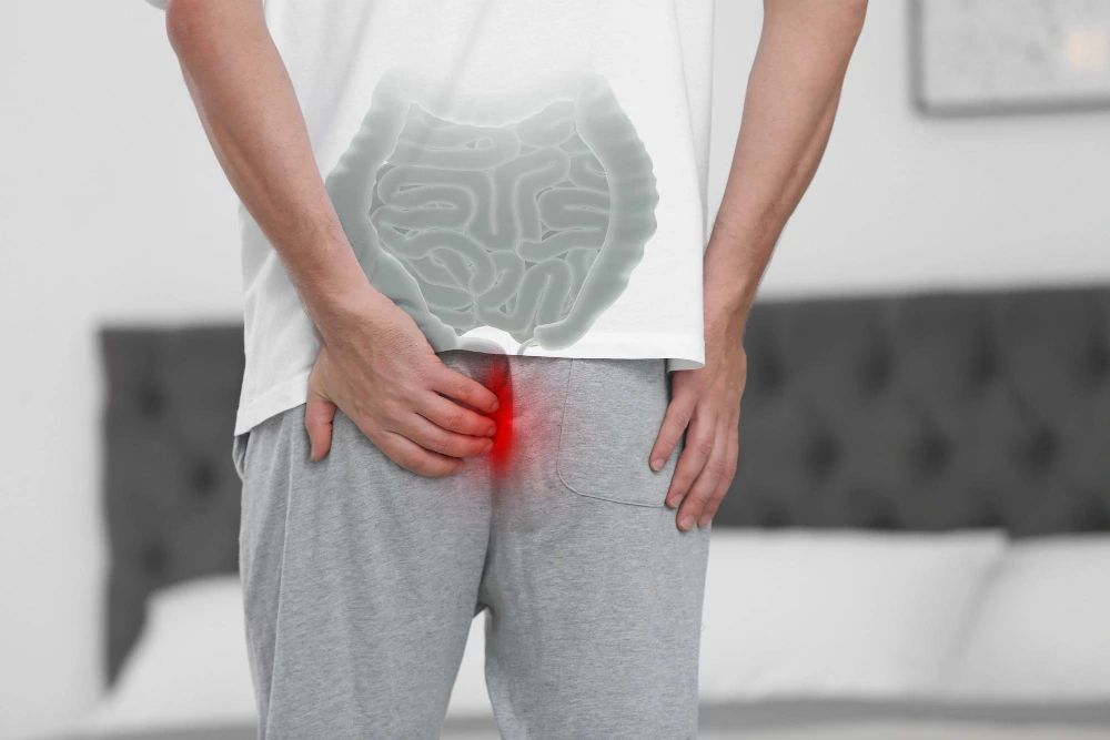
A hernia occurs when an internal organ or tissue pushes through a weak spot in the surrounding muscle or connective tissue. While hernias can appear in various parts of the body, they most commonly occur in the abdomen and groin region. The only effective treatment for a hernia is surgery, as hernias do not heal on their own and may worsen over time.
Modern medicine offers several types of hernia surgery, each designed to repair the defect, relieve pain, and prevent recurrence. Choosing the right type depends on the hernia’s location, severity, and the patient’s overall health.
Common Types of Hernia
Before understanding the types of hernia surgery, it’s essential to know the most common types of hernias:
| Type of Hernia | Location | Description |
| Inguinal Hernia | Groin area | Most common type, occurs when tissue pushes through a weak spot in the lower abdominal wall. |
| Femoral Hernia | Upper thigh | More common in women; occurs near the groin, below the inguinal ligament. |
| Umbilical Hernia | Belly button | Occurs when part of the intestine pushes through the abdominal wall near the navel. |
| Hiatal Hernia | Upper stomach | Occurs when part of the stomach pushes through the diaphragm into the chest cavity. |
| Incisional Hernia | Previous surgical site | Develops at the site of a previous incision due to weak tissue. |
| Epigastric Hernia | Upper abdomen | Occurs between the navel and the lower part of the rib cage. |
Types of Hernia Surgery
There are two main types of hernia surgery – open hernia repair and minimally invasive (laparoscopic or robotic-assisted) hernia repair. Each has its own benefits, risks, and recovery timelines.
1. Open Hernia Surgery (Open Herniorrhaphy)
Open hernia repair is the traditional approach used for many years. During this procedure, the surgeon makes a single incision over the hernia site to access the bulging tissue.
Procedure:
- The surgeon gently pushes the protruding tissue or organ back into its proper place.
- The weakened muscle wall is repaired, and a synthetic mesh may be placed to strengthen the area.
- The incision is then closed with sutures or staples.
Advantages:
- Suitable for large or complex hernias.
- Often performed under local or regional anesthesia.
- Shorter surgery time compared to laparoscopic methods.
Disadvantages:
- Larger incision and more postoperative discomfort.
- Longer recovery period.
- Slightly higher risk of infection and scarring.
Ideal For:
Patients with large hernias, recurrent hernias after laparoscopic surgery, or those not suitable for general anesthesia.
2. Laparoscopic Hernia Surgery
Laparoscopic hernia repair is a minimally invasive technique that uses small incisions and a laparoscope (a thin tube with a camera). It allows the surgeon to view and repair the hernia from inside the abdomen.
Procedure:
- The surgeon makes three to four small incisions.
- A laparoscope and surgical instruments are inserted.
- The hernia is repaired using a synthetic mesh, which is placed from inside to reinforce the abdominal wall.
- The incisions are closed with small stitches or surgical glue.
Advantages:
- Smaller incisions, leading to minimal scarring.
- Faster recovery and less postoperative pain.
- Lower risk of wound infection.
- Suitable for repairing bilateral or recurrent hernias.
Disadvantages:
- Requires general anesthesia.
- Slightly higher cost compared to open surgery.
- Not ideal for very large or complicated hernias.
Ideal For:
Patients who want a quick recovery, minimal pain, and smaller scars.
3. Robotic Hernia Surgery
Robotic hernia repair is an advanced version of laparoscopic surgery. It uses robotic technology to enhance precision and flexibility during the operation.
Procedure:
- Similar to laparoscopic surgery, but the surgeon operates robotic arms using a control console.
- The robot provides a 3D high-definition view, allowing better visualization of tissues.
- Mesh is placed to strengthen the weak spot.
Advantages:
- High precision and accuracy.
- Reduced blood loss.
- Quicker healing and minimal postoperative pain.
- Excellent for complex or recurrent hernias.
Disadvantages:
- Expensive compared to other methods.
- Requires specialized surgical expertise and facilities.
Ideal For:
Patients with complex, recurrent, or incisional hernias, and those seeking advanced, minimally invasive treatment.
4. Emergency Hernia Surgery
In rare cases, a hernia can become strangulated, meaning the blood supply to the trapped tissue is cut off. This is a medical emergency that requires immediate surgery.
Signs of a Strangulated Hernia:
- Severe pain and tenderness
- Redness or discoloration around the hernia site
- Nausea and vomiting
- Fever and rapid heart rate
Emergency hernia repair can be performed using open or laparoscopic methods, depending on the situation. The goal is to restore blood flow and repair the defect quickly to prevent complications.
Comparison Table: Types of Hernia Surgery
| Type | Anesthesia Used | Incision Size | Recovery Time | Scarring | Hospital Stay |
| Open Surgery | Local or general | Large | 4–6 weeks | Moderate | 1–2 days |
| Laparoscopic Surgery | General | Small (3–4 incisions) | 2–3 weeks | Minimal | Usually same day or overnight |
| Robotic Surgery | General | Small (3–4 incisions) | 1–2 weeks | Minimal | Usually same day |
| Emergency Surgery | General | Variable | Depends on condition | Variable | 2–4 days |
Which Type of Hernia Surgery is Best for You?
The choice between open, laparoscopic, or robotic surgery depends on several factors:
- Type and size of hernia: Large or complicated hernias may require open repair.
- Previous surgeries: Patients with previous hernia repairs might benefit from laparoscopic or robotic methods.
- Overall health: Laparoscopic and robotic surgeries are better for patients who can tolerate general anesthesia.
- Surgeon’s expertise: Availability of advanced technology and surgeon experience play a crucial role.
- Personal preference: Some patients prefer minimally invasive methods for quicker recovery and smaller scars.
A consultation with an experienced surgeon is essential to determine the most suitable surgical approach.
Recovery After Hernia Surgery
Recovery time varies depending on the type of hernia surgery performed. However, general recovery guidelines include:
Immediate Postoperative Period:
- Mild discomfort or pain around the incision site.
- Early movement is encouraged to prevent stiffness and promote blood circulation.
- Pain medication may be prescribed for the first few days.
First Few Weeks:
- Avoid heavy lifting, strenuous exercise, or sudden movements.
- Follow a light diet to prevent constipation and straining.
- Attend follow-up visits to ensure proper healing.
Long-Term Recovery Tips:
- Maintain a healthy weight.
- Avoid smoking, as it can delay healing.
- Practice good posture and body mechanics.
- Perform gentle strengthening exercises as advised by your doctor.
Risks and Complications
While hernia surgeries are generally safe, some potential risks include:
- Infection at the surgical site
- Bleeding or bruising
- Recurrence of hernia
- Chronic pain or nerve irritation
- Reaction to anesthesia
Choosing an experienced hernia surgeon and following postoperative care instructions significantly reduces these risks.
Cost of Hernia Surgery in India
The cost of hernia surgery varies depending on the type of procedure, hospital facilities, and location.
| Type of Surgery | Approximate Cost (₹) |
| Open Hernia Surgery | ₹40,000 – ₹70,000 |
| Laparoscopic Hernia Surgery | ₹70,000 – ₹1,20,000 |
| Robotic Hernia Surgery | ₹1,50,000 – ₹2,50,000 |
Insurance coverage is often available for hernia repair, depending on the policy terms.
When to See a Doctor
You should seek medical attention if you experience:
- A visible bulge in the abdomen or groin
- Pain or pressure at the hernia site
- Difficulty standing or lifting due to discomfort
- Nausea, vomiting, or changes in bowel habits
If the hernia becomes painful, discolored, or firm, it could indicate strangulation, a medical emergency that needs immediate surgery.
Conclusion
Choosing the right type of hernia surgery is crucial for effective and long-lasting results. With advancements in minimally invasive and robotic techniques, patients can expect faster recovery, minimal pain, and better outcomes than ever before.
At Kolekar Hospital, our experienced surgical team provides comprehensive diagnosis, personalized treatment plans, and advanced hernia surgery options tailored to your specific needs. Whether you require open, laparoscopic, or robotic-assisted repair, we ensure safe, effective, and patient-centered care from consultation to recovery.
![]()