Cardiology

When Is Angioplasty Needed? Symptoms, Indications and Treatment Options

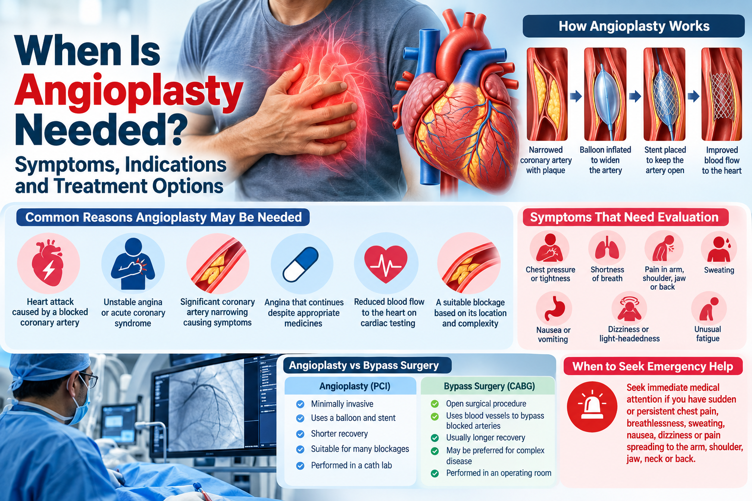
Angioplasty is a minimally invasive procedure used to improve blood flow through narrowed or blocked coronary arteries. It is commonly performed when fatty deposits, blood clots or other blockages reduce the supply of oxygen-rich blood to the heart muscle. During coronary angioplasty, a catheter is guided into the affected artery, usually with a small balloon used to widen the narrowed area. A stent is often placed to help keep the artery open.

If you have been diagnosed with coronary artery disease or have been advised to undergo angioplasty, you may wonder, “When is angioplasty needed?” The answer depends on several factors, including your symptoms, the severity and location of the blockage, whether you are having a heart attack, how well your symptoms respond to medicines, and your overall heart health.

Angioplasty is not automatically required for every person with a blocked or narrowed artery. A cardiologist evaluates the individual situation and considers medicines, lifestyle changes, angioplasty, stenting, or bypass surgery depending on the patient’s condition.

What Is Angioplasty?

Coronary angioplasty, also called percutaneous coronary intervention (PCI), is a procedure used to open a narrowed or blocked coronary artery.

The coronary arteries supply blood and oxygen to the heart muscle. Over time, plaque made up of cholesterol, fats and other substances can build up inside these arteries. This process, known as atherosclerosis, can narrow the arteries and reduce blood flow.

During angioplasty, a thin catheter is inserted through a blood vessel and guided toward the affected coronary artery. A small balloon at the tip of the catheter is inflated at the narrowed area to improve the opening. In most modern procedures, a stent is then placed to help maintain blood flow through the artery.

When Is Angioplasty Needed?

There is no single blockage percentage or symptom that automatically means angioplasty is necessary. The decision depends on the patient’s clinical condition and coronary anatomy.

Angioplasty may be considered in situations such as:

  • A heart attack caused by a blocked coronary artery
  • Unstable angina or acute coronary syndrome
  • Significant coronary artery narrowing causing troublesome symptoms
  • Angina that continues despite appropriate medicines
  • Reduced blood flow to the heart demonstrated by cardiac testing
  • A coronary blockage that is suitable for PCI based on its location and complexity

For stable coronary artery disease, treatment decisions are individualised. Some patients can manage their symptoms effectively with medication and lifestyle changes, while others may benefit from revascularisation. The choice between PCI, bypass surgery and medical treatment depends on factors such as symptoms, anatomy, disease complexity and overall health.

1. Angioplasty During a Heart Attack

One of the most important situations in which angioplasty may be needed is an acute heart attack.

A heart attack occurs when blood flow through a coronary artery is suddenly reduced or blocked, depriving part of the heart muscle of oxygen. In an appropriate patient, urgent PCI can reopen the affected artery and restore blood flow.

For certain heart attacks, particularly STEMI, primary PCI is an important emergency treatment when it can be performed promptly by an appropriate medical team.

Symptoms of a heart attack can include:

  • Chest pressure, tightness or discomfort
  • Pain or discomfort in the arm or shoulder
  • Pain spreading to the jaw, neck or back
  • Shortness of breath
  • Sweating
  • Nausea or vomiting
  • Light-headedness
  • Unusual weakness or fatigue

Women may also experience symptoms such as upper-back pressure, nausea, shortness of breath and unusual tiredness.

If these symptoms occur suddenly, seek emergency medical attention rather than waiting for them to disappear.

2. Angioplasty for Unstable Angina

Another important indication is unstable angina.

Unlike stable angina, which commonly occurs with physical activity and improves with rest, unstable angina may occur unexpectedly, including during rest. It can indicate a significant reduction in blood flow to the heart and may progress to a heart attack.

Depending on the results of the clinical assessment, ECG, blood tests and coronary angiography, treatment may include medicines, angioplasty with stenting, or coronary artery bypass surgery.

New, worsening or persistent chest discomfort should be treated as a medical emergency.

3. Angina That Does Not Improve With Medicines

People with stable angina are often initially treated with medicines and lifestyle changes. Medicines may include drugs that reduce the heart’s workload, improve blood flow, control cholesterol or reduce the risk of blood clots.

If angina continues despite appropriate medical treatment, a cardiologist may consider revascularisation, which can include angioplasty or bypass surgery depending on the coronary anatomy.

The purpose may be to improve blood flow and reduce symptoms that are limiting everyday activities. Angioplasty can decrease angina and improve the ability to perform physical activity when symptoms are caused by a suitable coronary blockage.

4. Significant Narrowing of a Coronary Artery

A coronary artery may become significantly narrowed because of plaque buildup. If the narrowing is affecting blood flow and is responsible for symptoms or evidence of myocardial ischaemia, angioplasty may be considered.

However, the decision is not based only on the percentage of narrowing seen on an angiogram. Doctors also consider:

  • Location of the blockage
  • Length and complexity of the lesion
  • Number of affected arteries
  • Symptoms
  • Results of other heart tests
  • Heart pumping function
  • Presence of diabetes or kidney disease
  • Previous heart procedures
  • Overall surgical and procedural risk

For complex coronary artery disease, a Heart Team may be involved in deciding whether PCI, bypass surgery or another treatment strategy is appropriate.

5. Evidence of Reduced Blood Flow to the Heart

Some people may have coronary artery narrowing without obvious chest pain. Doctors may identify reduced blood flow through tests such as a stress test or other cardiac imaging.

When test results indicate that a coronary blockage is clinically important, the cardiologist may discuss treatment options. Depending on the findings, treatment could include medication and lifestyle changes, angioplasty, or bypass surgery.

The goal is not simply to treat an image showing a blockage but to determine whether opening that artery is likely to benefit the patient.

Symptoms That May Indicate a Need for Cardiac Evaluation

Not every person who experiences chest discomfort needs angioplasty. However, symptoms suggestive of reduced blood flow to the heart should be evaluated.

Possible symptoms include:

SymptomWhat it may indicate
Chest pressure or tightnessPossible angina or another cardiac condition
BreathlessnessMay occur with reduced cardiac blood flow
Pain spreading to arm or shoulderPossible cardiac symptom
Jaw, neck or back discomfortCan occur with angina or heart attack
SweatingMay accompany acute coronary syndrome
Nausea or vomitingCan occur during a heart attack
Dizziness or light-headednessMay occur with cardiac problems
Unusual fatigueCan accompany heart disease, particularly in some women

These symptoms do not automatically mean that angioplasty is required. They indicate the need for appropriate medical evaluation.

How Do Doctors Decide Whether Angioplasty Is Needed?

Before recommending angioplasty, a cardiologist considers your symptoms, medical history, test results and coronary anatomy.

The evaluation may include:

Electrocardiogram

An ECG records the electrical activity of the heart and can help identify signs of a heart attack or other cardiac problems.

Blood Tests

Blood tests, including cardiac troponin testing when appropriate, can help identify heart muscle injury.

Echocardiogram

An echocardiogram uses ultrasound to assess the structure and pumping function of the heart.

Stress Testing

In selected patients, stress testing can help determine whether physical exertion is associated with reduced blood flow to the heart.

Coronary Angiography

Coronary angiography uses contrast dye and X-ray imaging to visualise the coronary arteries and identify narrowed or blocked areas. It can help doctors determine whether a blockage is suitable for PCI or whether another treatment approach is more appropriate.

Current chest-pain guidance emphasises structured risk assessment and appropriate cardiac testing rather than relying on symptoms alone.

Angioplasty vs Bypass Surgery

Angioplasty and coronary artery bypass grafting (CABG) are both methods of improving blood flow to the heart, but they work differently.

AngioplastyBypass Surgery
Minimally invasive catheter-based procedureOpen surgical procedure
Usually uses a balloon and stentUses blood vessels to create a new route around blocked arteries
Generally involves a shorter recoveryUsually involves a longer recovery
Suitable for many types of coronary blockagesMay be preferred for certain complex or extensive disease
Performed in a cardiac catheterisation laboratoryPerformed in an operating room

The choice between PCI and CABG depends on the patient’s coronary anatomy, disease complexity, symptoms, overall health and other factors. Some patients may benefit more from bypass surgery than angioplasty.

What Happens During Angioplasty?

Angioplasty is performed in a cardiac catheterisation laboratory.

A typical procedure involves:

  1. Preparing the patient and administering local anaesthesia.
  2. Inserting a thin catheter through a blood vessel, commonly in the wrist or sometimes the groin.
  3. Guiding the catheter to the coronary artery.
  4. Injecting contrast dye to identify the blockage.
  5. Positioning a small balloon at the narrowed area.
  6. Inflating the balloon to widen the artery.
  7. Placing a stent when appropriate.
  8. Confirming improved blood flow before completing the procedure.

The exact technique varies depending on the patient’s anatomy and clinical situation.

What Happens After Angioplasty?

After angioplasty, patients are monitored for a period of time. Some people may go home the same day or after a short hospital stay, while those treated during a heart attack or with other medical concerns may need longer hospitalisation.

Medicines are particularly important after stent placement. Depending on the type of stent and the patient’s circumstances, doctors commonly prescribe antiplatelet medicines to reduce the risk of blood clots forming inside the stent.

Patients should take prescribed medicines exactly as directed and should not stop antiplatelet medication without speaking with their cardiologist.

Cardiac rehabilitation, regular physical activity when medically appropriate, a heart-healthy diet, smoking cessation and control of blood pressure, cholesterol and diabetes are also important parts of long-term heart care.

Can Angioplasty Be Avoided?

In some patients, yes. Angioplasty is not necessary for every person with coronary artery disease.

Depending on the patient’s condition, treatment may involve:

  • Heart-healthy lifestyle changes
  • Cholesterol-lowering medicines
  • Blood pressure treatment
  • Antiplatelet medicines
  • Anti-anginal medicines
  • Diabetes management
  • Smoking cessation
  • Regular medical monitoring

For stable angina, medicines and lifestyle measures can often help control symptoms. If symptoms remain troublesome or the clinical situation changes, revascularisation may then be considered.

What Are the Risks of Angioplasty?

Angioplasty is commonly performed, but like any medical procedure, it carries potential risks.

These can include:

  • Bleeding or bruising at the catheter insertion site
  • Damage to a blood vessel
  • Allergic reaction to contrast dye
  • Abnormal heart rhythm
  • Blood clot formation
  • Re-narrowing of the treated artery
  • Heart attack or stroke
  • Kidney-related complications in susceptible patients

The individual risk depends on factors such as age, kidney function, overall health, the urgency of the procedure and the complexity of the coronary disease.

Your cardiologist should explain the expected benefits, risks and alternatives before a planned procedure.

When Should You Seek Emergency Medical Help?

Do not wait for a routine appointment if you develop sudden or persistent chest discomfort, particularly if it is accompanied by shortness of breath, sweating, nausea, dizziness or pain spreading to the arm, shoulder, jaw, neck or back.

Acute chest pain requires prompt medical assessment because it can be caused by a heart attack or another life-threatening condition.

Frequently Asked Questions About Angioplasty

When is angioplasty needed?

Angioplasty may be needed when a coronary artery is significantly narrowed or blocked and restoring blood flow is clinically appropriate. It is commonly used during certain heart attacks and may also be considered for unstable angina or persistent symptoms caused by suitable coronary artery disease.

Is angioplasty required for every blockage?

No. The presence of a blockage does not automatically mean that angioplasty is necessary. Doctors consider symptoms, blood-flow impairment, coronary anatomy, disease complexity and overall health before recommending treatment.

Is angioplasty the same as stent placement?

Not exactly. Angioplasty uses a balloon to widen a narrowed artery, while a stent is a small mesh tube placed inside the artery to help keep it open. In modern coronary PCI, angioplasty and stenting are commonly performed together.

How long does angioplasty take?

The procedure can vary in duration depending on the number and complexity of blockages and whether it is being performed as an emergency. Some procedures may take around 30 minutes, while more complex cases can take longer.

Can a person live normally after angioplasty?

Many people return to normal activities after recovery, but angioplasty does not remove the underlying tendency to develop coronary artery disease. Long-term medicines, follow-up care and heart-healthy lifestyle changes remain important.

Conclusion

Understanding when angioplasty is needed can help patients make informed decisions about their heart health. Angioplasty may be used to restore blood flow when a coronary artery is significantly narrowed or blocked, particularly during certain heart attacks and in selected patients with angina or other forms of coronary artery disease.

However, angioplasty is not automatically required for every blockage. A cardiologist considers the patient’s symptoms, test results, coronary anatomy, disease complexity and overall health before recommending PCI, medication, bypass surgery or another treatment approach.

If you experience sudden or persistent chest pressure, breathlessness, sweating, nausea, dizziness or pain spreading to the arm, shoulder, jaw, neck or back, seek emergency medical attention immediately. Early assessment and appropriate treatment can be critical when acute coronary syndrome or a heart attack is suspected.

Medical Disclaimer: This article is intended for general educational purposes only and should not replace professional medical advice, diagnosis or treatment. The decision to undergo angioplasty should be made after evaluation by a qualified cardiologist based on the individual patient’s clinical condition and test results.

Contact Details:

Kolekar Hospital & ICCU

Multispeciality Hospital in Chembur

Address: 2nd Floor, Omprakash Arcade, Approach to Ambedkar Garden Bus Stop Chembur Gaothan, Chembur 2nd & 3rd floor Omprakash Arcade, at Ambedkar garden, Chembur, Mumbai, Maharashtra 400071 

Phone: 02235083100

Website: kolekarhospital.com

Loading

Health

What Are the First Signs of Kidney Stones? Symptoms, Causes and Treatment

Kidney stones are hard, crystal-like deposits that develop inside the kidneys when certain minerals and salts become concentrated in the urine. They can vary in size, from tiny crystals that pass unnoticed to larger stones that can block the urinary tract and cause significant pain.

If you are wondering what are the first signs of kidney stones, one of the most common symptoms is pain in the back or side. However, kidney stones can cause several other symptoms, including changes in urination, blood in the urine, painful urination, nausea, vomiting and pain that moves toward the lower abdomen or groin.

Some kidney stones do not cause symptoms at all, particularly when they remain inside the kidney. Symptoms often begin when a stone moves into the ureter, the narrow tube that carries urine from the kidney to the bladder.

Recognising the early signs of kidney stones can help you seek medical attention before complications develop.

What Are the First Signs of Kidney Stones?

The first signs of kidney stones can vary depending on the size and location of the stone. A small stone may pass without causing noticeable symptoms, while a stone that becomes stuck in the ureter can cause sudden and severe discomfort.

Some of the early signs include:

  • Sharp pain in the side or back
  • Pain below the ribs
  • Pain that moves toward the lower abdomen or groin
  • Pain that comes and goes in waves
  • Blood in the urine
  • Burning or pain while urinating
  • Frequent urination
  • An urgent need to urinate
  • Nausea or vomiting
  • Cloudy or foul-smelling urine

It is important to remember that these symptoms can also occur with urinary tract infections and other conditions. Therefore, symptoms alone cannot confirm that you have a kidney stone.

1. Sharp Pain in the Back or Side

Pain is one of the most commonly recognised first signs of kidney stones. It usually occurs in the side or back, below the ribs, and may affect only one side of the body.

Kidney stone pain can be sudden and intense. It may also come in waves, becoming stronger and then temporarily improving.

As the stone moves through the urinary tract, the location of the pain may change. It can travel from the back or side toward the lower abdomen, pelvis or groin.

Unlike ordinary muscle pain, kidney stone pain may be difficult to relieve by changing your position. However, the severity and pattern can differ from person to person.

2. Pain That Moves Towards the Groin

A kidney stone travels from the kidney through the ureter toward the bladder. Because of this movement, the location of the pain can change.

Initially, you may feel pain in the side or back. As the stone moves downward, the discomfort may spread toward:

  • The lower abdomen
  • Pelvic region
  • Groin
  • Upper thigh

This changing pain pattern can be an important clue, especially when it occurs together with urinary symptoms.

3. Blood in the Urine

Another possible early sign of kidney stones is blood in the urine, medically known as hematuria.

The urine may appear:

  • Pink
  • Red
  • Brown
  • Tea-coloured

In some cases, the amount of blood may be too small to see with the naked eye and may only be detected during a urine test.

A stone can irritate the lining of the urinary tract as it moves, which may result in bleeding. However, blood in the urine can also be caused by urinary infections, kidney conditions and other medical problems. It should therefore be medically evaluated rather than automatically attributed to kidney stones.

4. Burning or Pain During Urination

As a kidney stone moves closer to the bladder, it can cause discomfort during urination.

You may experience:

  • Burning while passing urine
  • Sharp pain during urination
  • Pressure in the lower abdomen
  • Discomfort around the bladder or groin

Painful urination can also be caused by a urinary tract infection. If it occurs with fever, chills, cloudy urine or a strong urine odour, medical evaluation becomes particularly important.

5. Frequent or Urgent Urination

A kidney stone near the lower part of the urinary tract can make you feel that you need to urinate more frequently.

You may notice:

  • A frequent need to use the bathroom
  • A sudden or urgent need to urinate
  • Passing only a small amount of urine
  • Discomfort before or during urination

These symptoms can sometimes be mistaken for a urinary tract infection. A urine test and other investigations may be required to identify the actual cause.

6. Nausea and Vomiting

Kidney stones can cause nausea or vomiting, especially when the pain is severe.

The urinary system and digestive system are connected through shared nerve pathways. Intense pain caused by a stone can therefore trigger nausea or vomiting.

Nausea becomes more concerning when it is accompanied by:

  • Severe or persistent pain
  • Fever
  • Chills
  • Difficulty passing urine
  • Signs of dehydration

Repeated vomiting can also make it difficult to maintain adequate fluid intake, which may worsen dehydration.

7. Cloudy or Foul-Smelling Urine

Changes in the appearance or smell of urine can sometimes occur in people with kidney stones.

Urine may become cloudy or develop an unusually strong or unpleasant smell. However, these symptoms are also common with urinary tract infections.

If changes in urine occur along with fever, chills, pain or difficulty urinating, it is important to seek medical attention promptly.

8. Difficulty Passing Urine

A larger stone can become lodged in the urinary tract and interfere with the normal flow of urine.

You may notice that:

  • Urine flow becomes weaker
  • Only a small amount of urine is passed
  • You have difficulty starting urination
  • You feel unable to empty your bladder completely

Complete inability to pass urine can indicate a significant obstruction and requires urgent medical assessment.

Kidney Stone Symptoms at a Glance

SymptomWhat You May Experience
Back or side painSharp or severe pain below the ribs
Pain moving downwardDiscomfort spreading towards the abdomen or groin
Blood in urinePink, red or brown urine
Painful urinationBurning or discomfort while urinating
Frequent urinationNeeding to urinate more often
Urgent urinationSudden or persistent urge to urinate
NauseaFeeling sick, particularly with severe pain
VomitingMay occur during intense pain
Cloudy urineUrine appears less clear
Foul-smelling urineStrong or unusual urine odour
Fever or chillsMay indicate an associated infection
Difficulty urinatingReduced urine flow or inability to urinate

These symptoms are not exclusive to kidney stones, so proper medical evaluation is important.

Why Do Kidney Stones Form?

Kidney stones develop when certain substances in urine become highly concentrated and form crystals. These crystals can gradually combine and grow into stones.

Several factors can increase the likelihood of developing kidney stones, including:

  • Not drinking enough water
  • Excessive salt intake
  • A diet high in certain animal proteins
  • Obesity
  • A family history of kidney stones
  • Previous kidney stones
  • Certain digestive disorders
  • Recurrent urinary infections
  • Certain metabolic conditions
  • Some medications and supplements

There are different types of kidney stones, including calcium stones, uric acid stones, struvite stones and cystine stones. Identifying the type of stone can help doctors recommend appropriate preventive measures.

Can Kidney Stones Cause No Symptoms?

Yes. Kidney stones do not always cause noticeable symptoms.

A small stone may remain inside the kidney without causing pain. Some small stones may also pass through the urinary tract without the person realising it.

Symptoms are more likely when a stone moves into the ureter, becomes lodged, or interferes with the normal flow of urine.

Therefore, the absence of pain does not always mean that kidney stones are not present.

When Should You See a Doctor for Kidney Stones?

If you experience unexplained pain in your side or back, blood in your urine, painful urination or recurring urinary symptoms, it is advisable to consult a healthcare professional.

You should seek urgent medical attention if you experience:

  • Severe or unbearable pain
  • Fever or chills
  • Persistent vomiting
  • Blood in the urine
  • Difficulty passing urine
  • Inability to urinate
  • Severe pain that does not improve

Fever and chills are particularly important because they may indicate an infection. An infection occurring alongside urinary blockage can become serious and requires prompt treatment.

How Are Kidney Stones Diagnosed?

A doctor may diagnose kidney stones based on your symptoms, medical history, physical examination and diagnostic tests.

Depending on your symptoms, investigations may include:

Urine Tests

A urine test can identify blood, signs of infection and certain substances that may contribute to stone formation.

Blood Tests

Blood tests can help evaluate kidney function and check levels of substances such as calcium and uric acid.

Imaging Tests

Imaging can help determine whether a stone is present and identify its size and location. Depending on the patient’s age, symptoms and clinical condition, a doctor may recommend an ultrasound, CT scan or another appropriate imaging test.

How Are Kidney Stones Treated?

Treatment depends on several factors, including the size and location of the stone, whether it is causing an obstruction, and whether an infection is present.

Small kidney stones may sometimes pass naturally. Doctors may recommend adequate fluid intake, pain management and monitoring, depending on the individual situation.

For larger stones or stones that do not pass naturally, treatment may include procedures such as:

  • Shock wave lithotripsy
  • Ureteroscopy
  • Other minimally invasive stone removal procedures
  • Surgical treatment in selected cases

A urologist can determine the most suitable treatment based on the individual patient’s condition.

How Can Kidney Stones Be Prevented?

Drinking enough water is one of the most important measures for reducing the risk of kidney stones. Adequate hydration helps dilute substances in the urine that can form crystals.

Other preventive measures may include:

  • Drinking sufficient water throughout the day
  • Reducing excessive salt intake
  • Maintaining a balanced diet
  • Maintaining a healthy body weight
  • Following medical advice regarding dietary calcium and oxalate
  • Managing conditions that increase stone risk
  • Following prescribed preventive medicines when required

If you have had kidney stones previously, your doctor may recommend additional tests to determine why they formed and how future stones can be prevented.

Frequently Asked Questions About Kidney Stones

What are the first signs of kidney stones?

The first signs of kidney stones can include sharp pain in the side or back, pain that moves towards the lower abdomen or groin, blood in the urine, painful urination, frequent urination, nausea and vomiting. Some people may have no symptoms until the stone moves into the ureter.

Where does kidney stone pain usually start?

Kidney stone pain commonly starts in the side or back below the ribs. As the stone moves through the urinary tract, the pain may travel towards the lower abdomen or groin.

Can kidney stones cause blood in urine?

Yes. A kidney stone can irritate the urinary tract and cause blood in the urine. The urine may appear pink, red or brown, although blood may sometimes only be detected through laboratory testing.

Can kidney stones cause frequent urination?

Yes. A stone, particularly one that has moved closer to the bladder, may cause a frequent or urgent need to urinate.

Can kidney stones go away on their own?

Some smaller kidney stones can pass naturally without a procedure. Whether a stone will pass depends on factors such as its size and location. A doctor can determine whether monitoring is appropriate or whether treatment is necessary.

Are kidney stones dangerous?

Many kidney stones can be treated successfully, but a stone that blocks urine flow or occurs together with an infection can become a serious medical problem. Prompt medical evaluation is important when severe symptoms occur.

Conclusion

Knowing what are the first signs of kidney stones can help you recognise symptoms and seek medical attention at the right time. Sharp pain in the side or back, pain that moves towards the lower abdomen or groin, blood in the urine, painful urination, frequent urination, nausea and vomiting are some of the symptoms that may occur when a kidney stone moves through the urinary tract.

However, these symptoms can also occur with other urinary or abdominal conditions. Severe pain, fever, chills, repeated vomiting or difficulty passing urine should not be ignored.

If you experience symptoms that may indicate kidney stones, consult a qualified healthcare professional for an accurate diagnosis. Early evaluation can help determine the size and location of the stone, identify possible complications and guide appropriate treatment.

Medical Disclaimer: This blog is intended for general educational purposes and should not replace professional medical advice, diagnosis or treatment. If you experience severe pain, fever, chills, persistent vomiting, blood in the urine or difficulty urinating, seek prompt medical attention.

Contact Details:

Kolekar Hospital & ICCU

Multispeciality Hospital in Chembur

Address: 2nd Floor, Omprakash Arcade, Approach to Ambedkar Garden Bus Stop Chembur Gaothan, Chembur 2nd & 3rd floor Omprakash Arcade, at Ambedkar garden, Chembur, Mumbai, Maharashtra 400071 

Phone: 02235083100

Website: kolekarhospital.com

Loading

Cardiology

What Causes Chest Pain in a Woman? Causes, Symptoms and When to Seek Medical Help

Chest pain can be worrying, especially when it occurs suddenly or without an obvious reason. If you are wondering what causes chest pain in a woman, it is important to understand that there can be several possible causes. While chest pain is often associated with heart problems, it can also result from acid reflux, muscle strain, anxiety, lung conditions, hormonal changes, or breast-related problems.

Women may also experience heart-related conditions differently from men. Instead of severe or crushing chest pain, some women may experience shortness of breath, unusual tiredness, nausea, dizziness, sweating, or discomfort in the back, shoulder, arm, neck, or jaw.

Because chest pain can sometimes indicate a medical emergency, it should never be ignored, particularly when it is new, severe, persistent, or accompanied by other concerning symptoms.

What Causes Chest Pain in a Woman?

There is no single answer to what causes chest pain in a woman because chest discomfort can originate from different organs and tissues. The heart, lungs, digestive system, muscles, nerves, and breast tissue can all contribute to pain felt in or around the chest.

Some common causes include:

  • Heart conditions such as angina or heart attack
  • Acid reflux and indigestion
  • Muscle strain or chest-wall inflammation
  • Anxiety and panic attacks
  • Lung infections and other respiratory conditions
  • Inflammation around the heart
  • Breast-related conditions
  • Hormonal changes

The nature of the pain, when it occurs, how long it lasts, and whether it is associated with other symptoms can provide useful information. However, these symptoms alone cannot reliably determine the cause. A healthcare professional may need to perform an examination and appropriate tests.

Heart-Related Causes of Chest Pain in Women

One of the most important causes of chest pain to consider is a problem involving the heart. Conditions such as coronary artery disease, angina, and heart attack can cause chest discomfort.

Angina

Angina occurs when the heart muscle does not receive enough oxygen-rich blood. It may feel like pressure, tightness, squeezing, heaviness, or discomfort in the chest.

The discomfort may sometimes spread to the:

  • Arm or shoulder
  • Back
  • Neck
  • Jaw

Women may experience symptoms such as shortness of breath, nausea, or discomfort in areas other than the chest. Angina may occur during physical activity or emotional stress and may improve with rest, depending on the type.

Heart Attack

A heart attack occurs when blood flow to part of the heart becomes significantly reduced or blocked. It requires immediate medical attention.

Chest pressure or discomfort is an important warning sign, but women may also experience symptoms such as:

  • Shortness of breath
  • Nausea or vomiting
  • Cold sweating
  • Dizziness
  • Unusual tiredness
  • Pain or discomfort in the back, shoulder, arm, neck, or jaw

Some women may have relatively mild symptoms, and symptoms can sometimes develop gradually. This is why unexplained chest discomfort should not automatically be dismissed as acidity, gas, or stress.

Can Acid Reflux Cause Chest Pain in Women?

Yes. Acid reflux and indigestion are common causes of chest discomfort. Gastroesophageal reflux disease (GERD) occurs when stomach contents flow back into the oesophagus, causing irritation.

Acid reflux-related chest discomfort may feel like burning behind the breastbone and may be accompanied by:

  • Heartburn
  • A sour or bitter taste in the mouth
  • Burping
  • Bloating
  • Discomfort after meals

Symptoms may become worse after eating or when lying down.

However, heartburn and heart-related chest pain can sometimes feel similar. Therefore, new or unexplained chest pain should not automatically be assumed to be caused by acidity.

Muscle Strain and Chest Wall Pain

Another possible explanation for chest pain is a problem involving the muscles, ribs, or chest wall.

Muscle-related chest pain can occur after:

  • Exercise or weightlifting
  • Repetitive upper-body movements
  • Coughing for a prolonged period
  • An injury
  • Sudden physical exertion
  • Poor posture

This type of pain may become more noticeable when moving the upper body or touching the affected area.

Although muscle strain is generally less serious than many heart-related causes, it is still important to seek medical advice when the cause of chest pain is uncertain.

Can Anxiety Cause Chest Pain in Women?

Anxiety and panic attacks can cause physical symptoms, including chest discomfort. During a panic attack, a person may experience chest tightness along with rapid heartbeat, sweating, dizziness, trembling, or shortness of breath.

Stress can also cause muscle tension, which may contribute to discomfort around the chest and shoulders.

However, chest pain should not automatically be attributed to anxiety. Heart, lung, and other medical conditions can produce similar symptoms. If the pain is new, severe, persistent, or associated with other warning signs, medical evaluation is important.

Lung Conditions That Can Cause Chest Pain

Certain respiratory conditions can also cause chest pain. Depending on the condition, the pain may become worse when coughing or taking a deep breath.

Possible causes include:

  • Pneumonia
  • Pleurisy
  • Respiratory infections
  • Pulmonary embolism
  • Certain chronic lung conditions

Chest pain associated with a lung problem may occur together with cough, fever, difficulty breathing, or a rapid heartbeat.

A pulmonary embolism, which occurs when a blood clot blocks an artery in the lungs, can be life-threatening. Sudden chest pain combined with significant shortness of breath, rapid heartbeat, coughing up blood, or fainting requires immediate emergency medical attention.

Pericarditis and Other Heart-Related Conditions

Pericarditis is inflammation of the thin sac surrounding the heart. It can cause sharp or stabbing chest pain. The discomfort may become worse with deep breathing, coughing, or lying down.

Other heart-related conditions can also cause chest discomfort, which is why identifying the exact cause requires proper medical assessment.

If chest pain is persistent or accompanied by breathlessness, dizziness, sweating, fainting, or pain spreading to other areas of the body, urgent medical care may be required.

Breast-Related Causes of Chest Discomfort

Sometimes women may describe pain around the chest when the actual source is breast tissue.

Breast pain or tenderness can occur due to:

  • Hormonal changes during the menstrual cycle
  • Pregnancy
  • Breast cysts
  • Breast infections
  • Injury
  • Poor breast support

Breast pain alone is not usually a sign of breast cancer. However, persistent breast pain accompanied by a new lump, skin changes, nipple changes, or unusual discharge should be evaluated by a healthcare professional.

Hormonal Changes and Chest or Breast Pain

Hormonal fluctuations can cause breast tenderness or discomfort in some women. Changes in oestrogen and progesterone levels during the menstrual cycle may make the breasts feel swollen, sensitive, or painful.

Similar symptoms may occur during pregnancy, perimenopause, and menopause.

Even when hormonal changes are suspected, persistent or unusual chest pain should not automatically be considered hormonal. A healthcare professional can determine whether further evaluation is necessary.

Chest Pain in Women: Understanding the Possible Causes

Possible CauseSymptoms or Features
Heart-related conditionsPressure, tightness, heaviness, shortness of breath or discomfort spreading to the upper body
Acid refluxBurning sensation, heartburn, sour taste, discomfort after meals
Muscle strainPain associated with movement, exercise, coughing, or injury
Anxiety or panicChest tightness, rapid heartbeat, sweating, dizziness
Lung conditionsPain with breathing, cough, fever, or shortness of breath
PericarditisSharp chest pain that may worsen with deep breathing or lying down
Breast-related causesLocalised breast tenderness or pain
Hormonal changesBreast tenderness or discomfort related to menstrual or hormonal changes

These symptoms are general patterns and cannot be used to diagnose the cause of chest pain.

Why Chest Pain Symptoms Can Be Different in Women

One important reason women should take chest discomfort seriously is that heart-related symptoms may not always appear in the same way as they are traditionally described.

While chest pressure or discomfort is an important symptom, some women may experience:

  • Unusual or unexplained fatigue
  • Shortness of breath
  • Nausea
  • Dizziness
  • Cold sweating
  • Back or shoulder discomfort
  • Jaw or neck pain
  • Discomfort in one or both arms

These symptoms can occur with or without significant chest pain.

For this reason, women should pay attention to unusual combinations of symptoms, particularly if they develop suddenly or occur during physical activity.

When Should a Woman Seek Emergency Medical Care for Chest Pain?

Chest pain requires urgent medical attention when it is sudden, severe, persistent, or associated with symptoms that could indicate a heart or lung emergency.

Seek emergency medical help if chest pain occurs with:

  • Severe pressure, squeezing, or heaviness
  • Difficulty breathing
  • Cold sweating
  • Nausea or vomiting
  • Fainting or severe dizziness
  • Rapid or irregular heartbeat
  • Pain spreading to the arm, shoulder, back, neck, or jaw
  • Sudden weakness or unusual extreme fatigue

Do not wait for symptoms to become severe before seeking help if you suspect a medical emergency.

How Is Chest Pain Diagnosed?

When you visit a doctor for chest pain, the evaluation may begin with questions about the location, duration, severity, and nature of the pain. Your medical history and risk factors are also important.

Depending on the symptoms, your doctor may recommend tests such as:

  • Electrocardiogram (ECG)
  • Blood tests
  • Blood pressure and oxygen-level measurement
  • Echocardiogram
  • Chest X-ray
  • Stress testing
  • CT scans
  • Coronary angiography

The tests required depend on the suspected cause and whether the symptoms suggest an emergency.

Risk Factors for Heart-Related Chest Pain in Women

Certain health and lifestyle factors can increase the risk of cardiovascular disease. These may include:

  • High blood pressure
  • Diabetes
  • High cholesterol
  • Smoking
  • Obesity
  • Physical inactivity
  • Family history of heart disease
  • Increasing age
  • Certain pregnancy-related health conditions

Regular health check-ups can help identify and manage several of these risk factors.

How Can Women Reduce Their Risk of Heart Disease?

Although not every cause of chest pain can be prevented, maintaining cardiovascular health can help reduce the risk of heart disease.

Helpful measures include:

  • Eating a balanced diet
  • Staying physically active
  • Avoiding smoking and tobacco
  • Maintaining a healthy weight
  • Managing blood pressure and cholesterol
  • Controlling diabetes if present
  • Getting adequate sleep
  • Managing ongoing stress
  • Attending regular health check-ups

Women with known risk factors should discuss appropriate preventive measures with their healthcare provider.

Frequently Asked Questions About Chest Pain in Women

What causes chest pain in a woman most commonly?

Chest pain in women can have several causes, including acid reflux, muscle strain, anxiety, respiratory conditions, breast-related discomfort, and heart-related conditions. The exact cause depends on the individual’s symptoms and health history.

Can gas cause chest pain in women?

Gas and indigestion can cause discomfort in the upper abdomen or chest. However, heart-related pain can sometimes feel similar to indigestion. New or unexplained chest pain should therefore be assessed rather than automatically attributed to gas.

Can stress cause chest pain in women?

Yes. Stress and anxiety can contribute to chest tightness, muscle tension, rapid heartbeat, and other physical symptoms. However, it is important to rule out medical causes when chest pain is new, severe, persistent, or accompanied by concerning symptoms.

Is chest pain always a sign of a heart problem?

No. Chest pain can be caused by digestive, muscular, respiratory, hormonal, breast-related, and other conditions. However, because some heart-related causes can be serious or life-threatening, unexplained chest pain should be taken seriously.

When should chest pain be treated as an emergency?

Seek immediate medical attention if chest pain is severe, persistent, or accompanied by difficulty breathing, sweating, nausea, fainting, dizziness, or discomfort spreading to the arm, shoulder, back, neck, or jaw.

Conclusion

Understanding what causes chest pain in a woman is important because chest discomfort can have many different causes. Acid reflux, muscle strain, anxiety, hormonal changes, breast-related conditions, lung problems, and heart conditions can all produce symptoms in or around the chest.

While some causes may be relatively minor, heart attacks and certain lung conditions can be life-threatening. Women may also experience heart-related problems with symptoms such as unusual fatigue, nausea, shortness of breath, dizziness, sweating, or discomfort in the back, shoulder, arm, neck, or jaw.

Therefore, new, unexplained, severe, or persistent chest pain should not be ignored. If symptoms suggest a medical emergency, seek immediate medical attention. For recurring or unexplained symptoms, consult a qualified healthcare professional for appropriate evaluation and treatment.

Medical Disclaimer: This article is intended for general educational purposes only and should not be considered a substitute for professional medical advice, diagnosis, or treatment. If you are experiencing severe or concerning chest pain, seek emergency medical care immediately.

Contact Details:

Kolekar Hospital & ICCU

Multispeciality Hospital in Chembur

Address: 2nd Floor, Omprakash Arcade, Approach to Ambedkar Garden Bus Stop Chembur Gaothan, Chembur 2nd & 3rd floor Omprakash Arcade, at Ambedkar garden, Chembur, Mumbai, Maharashtra 400071 

Phone: 02235083100

Website: kolekarhospital.com

Loading

Uncategorized

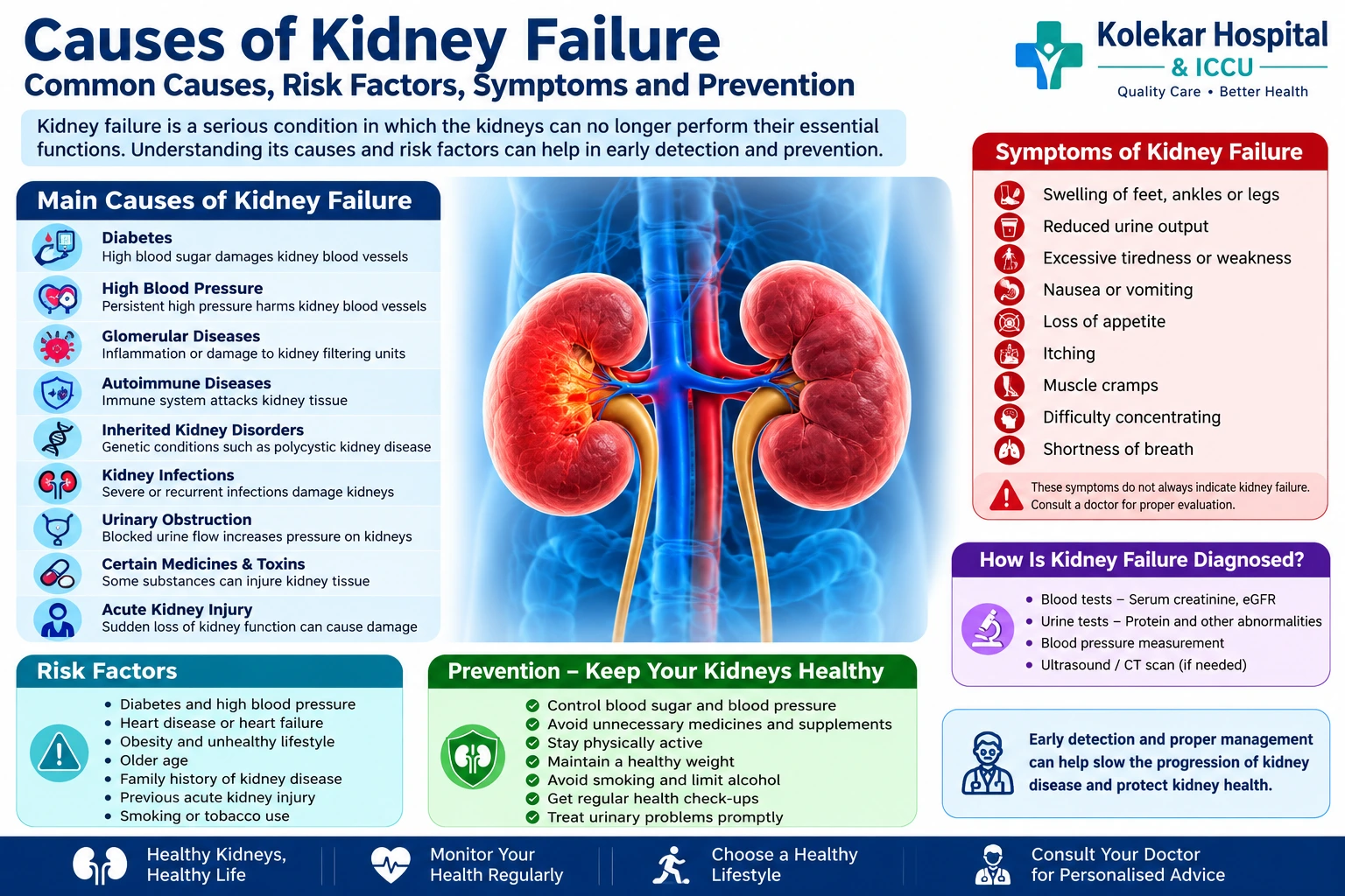
Causes of Kidney Failure: Common Causes, Risk Factors, Symptoms and Prevention

Kidney failure is a serious medical condition in which the kidneys can no longer perform their essential functions adequately. Healthy kidneys filter waste products and excess fluid from the blood, maintain the body’s electrolyte and mineral balance, help regulate blood pressure, support red blood cell production and contribute to healthy bones.

Kidney failure can develop gradually over several years because of chronic kidney disease, or it can occur suddenly as a result of acute kidney injury. Understanding the causes of kidney failure is important because many of the conditions that damage the kidneys can be detected and managed before permanent kidney damage occurs.

The two most common causes of chronic kidney disease and kidney failure are diabetes and high blood pressure. However, several other conditions, including autoimmune diseases, inherited kidney disorders, infections, urinary tract obstruction, certain medications and toxins, can also cause progressive kidney damage.

What Is Kidney Failure?

Kidney failure occurs when kidney function becomes severely reduced and the kidneys are unable to adequately remove waste, control fluid levels and maintain the body’s internal chemical balance.

Kidney failure may develop through:

  • Chronic kidney disease (CKD): A gradual decline in kidney function over months or years.
  • Acute kidney injury (AKI): A sudden decrease in kidney function that can occur over hours or days.
  • Acute kidney injury on chronic kidney disease: A sudden deterioration in someone who already has chronic kidney damage.

Kidney failure is generally associated with kidney function falling to a very low level. The National Institute of Diabetes and Digestive and Kidney Diseases (NIDDK) describes kidney failure as kidney function below approximately 15% of normal.

When kidney function becomes severely impaired, waste products and excess fluid can accumulate in the body. Depending on the severity and cause, treatment may include medicines, dietary changes, dialysis or kidney transplantation.

What Are the Main Causes of Kidney Failure?

The causes of kidney failure can be broadly divided into diseases that gradually damage the kidneys and conditions that cause sudden kidney injury.

CauseHow It Can Damage the Kidneys
DiabetesHigh blood sugar can damage kidney blood vessels and filtering units
High blood pressurePersistent pressure damages kidney blood vessels and filtration structures
Glomerular diseasesInflammation or damage affects the kidney’s filtering units
Autoimmune diseasesThe immune system can attack kidney tissue
Polycystic kidney diseaseGenetic cysts progressively damage normal kidney tissue
Kidney infectionsSevere or recurrent infections may cause kidney damage
Urinary obstructionBlocked urine flow can increase pressure and damage the kidneys
Kidney stonesRepeated obstruction or complications can affect kidney function
Certain medicines and toxinsSome substances can directly injure kidney tissue
Acute kidney injurySudden loss of kidney function can sometimes result in permanent damage

1. Diabetes Is a Major Cause of Kidney Failure

Diabetes is one of the most important causes of chronic kidney disease and kidney failure. Persistently high blood glucose can damage the small blood vessels and filtering structures within the kidneys.

The kidneys contain millions of microscopic filtering units called nephrons. Over time, elevated blood sugar can damage these structures, allowing proteins such as albumin to leak into the urine while reducing the kidneys’ ability to filter waste effectively.

This condition is commonly referred to as diabetic kidney disease.

People with diabetes may not experience obvious kidney symptoms in the early stages. This makes regular monitoring particularly important.

Diabetes-related kidney damage can be reduced by:

  • Maintaining blood glucose within the target range recommended by a doctor
  • Monitoring blood pressure
  • Taking prescribed medicines consistently
  • Getting regular kidney function and urine tests
  • Following an appropriate diet
  • Maintaining a healthy weight
  • Avoiding tobacco products

Early detection is important because kidney damage may progress silently before noticeable symptoms develop.

2. High Blood Pressure and Kidney Failure

High blood pressure, or hypertension, is another leading cause of kidney damage.

Blood vessels inside the kidneys are delicate and designed to filter large volumes of blood. Persistently elevated blood pressure can damage these blood vessels and impair the kidney’s filtration system. As kidney function declines, the kidneys may also become less effective at controlling blood pressure, creating a cycle in which hypertension and kidney disease can worsen each other.

Uncontrolled hypertension over many years can therefore contribute to chronic kidney disease and eventually kidney failure.

People with high blood pressure should follow their doctor’s recommendations regarding:

  • Regular blood pressure monitoring
  • Prescribed antihypertensive medicines
  • Limiting excessive salt intake
  • Maintaining a healthy weight
  • Regular physical activity
  • Managing diabetes and cholesterol
  • Avoiding smoking

3. Glomerular Diseases

The glomeruli are tiny filtering structures within the kidneys. Diseases that damage these structures are collectively known as glomerular diseases.

Examples include:

  • Glomerulonephritis
  • IgA nephropathy
  • Membranous nephropathy
  • Focal segmental glomerulosclerosis
  • IgA vasculitis

Inflammation or structural damage to the glomeruli can cause blood or protein to appear in the urine and gradually reduce kidney function. Some glomerular diseases develop because of immune-system abnormalities, while others may be associated with infections or other medical conditions.

If left untreated or if severe, some of these conditions can progress to advanced kidney disease.

4. Autoimmune Diseases

Autoimmune diseases can also be among the causes of kidney failure.

In autoimmune disorders, the immune system mistakenly attacks the body’s own tissues. One important example is lupus nephritis, in which systemic lupus erythematosus affects the kidneys.

Other immune-mediated diseases can also affect the kidney’s filtering structures.

Patients with autoimmune disease may require regular monitoring of:

  • Blood pressure
  • Serum creatinine and estimated glomerular filtration rate (eGFR)
  • Urine protein
  • Urine blood
  • Other disease-specific markers

Early treatment of kidney involvement may help slow the progression of kidney damage.

5. Polycystic Kidney Disease and Other Inherited Conditions

Some kidney diseases are caused by genetic changes that can be inherited within families.

Polycystic kidney disease (PKD) is one of the better-known inherited kidney disorders. It causes multiple cysts to develop in the kidneys. Over time, these cysts can enlarge and interfere with normal kidney tissue.

Other inherited disorders can also impair kidney function.

A family history of kidney disease or kidney failure can increase a person’s risk. People with a strong family history should discuss appropriate screening with a healthcare professional.

6. Kidney Infections and Recurrent Urinary Tract Infections

A urinary tract infection (UTI) usually affects the bladder or lower urinary tract. However, an infection can sometimes spread to the kidneys and cause pyelonephritis, or kidney infection.

Repeated, severe or inadequately treated kidney infections can potentially cause scarring and long-term kidney damage.

The risk can be particularly important in people who have:

  • Urinary tract abnormalities
  • Kidney stones
  • Urinary obstruction
  • Conditions affecting bladder emptying
  • Recurrent infections

Persistent symptoms such as fever, chills, painful urination or pain in the side or back should be medically evaluated rather than repeatedly self-treated.

7. Kidney Stones and Urinary Tract Obstruction

Kidney stones can sometimes block the normal flow of urine. Other causes of urinary obstruction include:

  • Enlarged prostate
  • Tumours
  • Ureteral narrowing or scarring
  • Congenital urinary tract abnormalities
  • Severe urinary retention
  • Large or obstructing stones

When urine cannot flow normally, pressure can build up within the urinary system. Prolonged obstruction can damage the kidneys and may contribute to loss of kidney function.

This is why persistent urinary difficulty, reduced urine output or recurrent urinary obstruction should not be ignored.

8. Acute Kidney Injury

Not all kidney failure develops slowly. Acute kidney injury (AKI) is a sudden reduction in kidney function that can develop over a short period.

AKI may occur because of:

  • Severe dehydration
  • Major blood loss
  • Very low blood pressure
  • Severe infections or sepsis
  • Certain medicines
  • Exposure to kidney-toxic substances
  • Severe illness
  • Urinary obstruction
  • Major surgery or trauma

NIDDK notes that AKI can result from conditions such as sepsis, decreased blood pressure and kidney injury caused by drugs or toxins.

Some people recover substantially after AKI, particularly when the underlying cause is identified and treated promptly. However, severe AKI can sometimes result in persistent kidney damage or increase the risk of developing chronic kidney disease.

9. Certain Medicines and Toxic Substances

Some medicines can affect kidney function, particularly when used inappropriately, at excessive doses or in people who already have kidney problems.

Examples may include certain:

  • Pain-relieving medicines
  • Antibiotics
  • Chemotherapy medicines
  • Other medicines that can be nephrotoxic in particular circumstances

The National Kidney Foundation also identifies excessive use of non-steroidal anti-inflammatory drugs (NSAIDs), certain drugs, contrast substances and environmental toxins among potential contributors to kidney damage.

This does not mean patients should stop prescribed medicines on their own. Instead, people with kidney disease should tell their doctor about all prescription medicines, over-the-counter medicines and supplements they use.

10. Severe Infections and Other Serious Medical Conditions

Severe systemic infections can cause widespread inflammation, low blood pressure and reduced blood flow to the kidneys. This may result in acute kidney injury.

Conditions such as haemolytic uremic syndrome (HUS) and thrombotic thrombocytopenic purpura (TTP) can also affect kidney function. Certain cancers and blood-related conditions, including multiple myeloma, may contribute to chronic kidney disease in some patients.

These conditions require specialised medical evaluation because the appropriate treatment depends on the underlying cause.

Risk Factors That Can Increase the Chance of Kidney Failure

Having a risk factor does not necessarily mean that a person will develop kidney failure. However, several factors can increase the likelihood of kidney disease.

Common risk factors include:

  • Diabetes
  • High blood pressure
  • Heart disease or heart failure
  • Obesity
  • Older age
  • Family history of kidney disease or kidney failure
  • Previous acute kidney injury
  • Smoking or tobacco use
  • Certain inherited disorders
  • Recurrent urinary tract problems
  • Long-term exposure to kidney-toxic medicines or substances

Diabetes and hypertension remain the most important common causes of chronic kidney disease.

What Are the Symptoms of Kidney Failure?

Kidney disease can be difficult to recognise in its early stages because symptoms may be absent or nonspecific.

As kidney function becomes severely impaired, symptoms may include:

  • Swelling of the feet, ankles or legs
  • Reduced urine output
  • Excessive tiredness or weakness
  • Nausea or vomiting
  • Loss of appetite
  • Itching
  • Muscle cramps
  • Difficulty concentrating
  • Sleep problems
  • Shortness of breath
  • Changes in urination

These symptoms do not necessarily mean that a person has kidney failure. They can occur with several other medical conditions, so proper evaluation is important.

How Is Kidney Failure Diagnosed?

Doctors use a combination of medical history, physical examination, blood tests, urine tests and, when required, imaging studies.

Common investigations include:

TestWhat It Helps Assess
Serum creatinineKidney filtration and overall kidney function
eGFREstimated level of kidney filtration
Urine albumin-creatinine ratioProtein leakage through the kidneys
UrinalysisBlood, protein and other abnormalities in urine
Blood pressure measurementIdentifies an important kidney disease risk factor
Ultrasound/CT or other imagingMay identify obstruction, stones or structural abnormalities

The National Kidney Foundation identifies eGFR and urine albumin-creatinine ratio as important tests for assessing kidney health.

Can Kidney Failure Be Prevented?

Not every case of kidney failure can be prevented, particularly when it results from inherited or unavoidable medical conditions. However, many important risk factors can be controlled.

Steps that can support kidney health include:

  1. Control blood sugar: People with diabetes should follow their treatment plan and monitor glucose as recommended.
  2. Control blood pressure: Regular monitoring and appropriate treatment can reduce kidney damage.
  3. Avoid unnecessary medicines: Do not regularly take painkillers or supplements without discussing their safety with a healthcare professional.
  4. Stay physically active: Regular exercise supports cardiovascular and metabolic health.
  5. Maintain a healthy weight: Obesity is associated with increased kidney disease risk.
  6. Avoid smoking: Tobacco use can adversely affect blood vessels and overall kidney health.
  7. Get regular check-ups: People at higher risk should discuss kidney screening with their doctor.
  8. Treat urinary problems promptly: Recurrent infections, stones or difficulty passing urine should be evaluated.
  9. Follow medical advice after acute kidney injury: Previous AKI can increase the risk of future chronic kidney disease.

When Should You See a Kidney Specialist?

You should consider medical evaluation if you have persistent changes in urination, unexplained swelling, blood or protein in the urine, consistently high blood pressure, abnormal kidney function tests or a history of conditions that can damage the kidneys.

People with diabetes, hypertension, cardiovascular disease, a family history of kidney failure or previous acute kidney injury may also benefit from regular kidney function monitoring.

Early assessment is particularly important because kidney disease can progress without obvious symptoms.

Frequently Asked Questions About the Causes of Kidney Failure

What are the most common causes of kidney failure?

The two major causes are diabetes and high blood pressure. Other causes include glomerular diseases, autoimmune disorders, inherited kidney diseases, infections, urinary obstruction, kidney stones, certain medicines and acute kidney injury.

Can high blood pressure cause kidney failure?

Yes. Long-standing, poorly controlled high blood pressure can damage the blood vessels and filtering structures of the kidneys. Kidney disease can also worsen blood pressure, creating a cycle of progressive damage.

Can diabetes lead to kidney failure?

Yes. Persistently high blood sugar can damage the kidney’s filtering system. Over time, this can result in diabetic kidney disease and, in severe cases, kidney failure.

Can kidney failure happen suddenly?

Yes. A sudden decline in kidney function is called acute kidney injury. Severe dehydration, low blood pressure, sepsis, certain medicines, toxins and urinary obstruction are among the potential causes.

Can kidney failure be reversed?

It depends on the cause. Some cases of acute kidney injury can improve substantially when the underlying problem is treated promptly. Advanced chronic kidney failure generally represents permanent loss of kidney function and may require long-term kidney replacement treatment such as dialysis or transplantation.

Can kidney failure occur without symptoms?

Yes. Chronic kidney disease can remain unnoticed for a long time. This is one reason why people with diabetes, high blood pressure and other risk factors should undergo appropriate kidney health checks.

Conclusion

Understanding the causes of kidney failure is an important part of protecting long-term kidney health. Diabetes and high blood pressure are the leading causes, but kidney failure can also result from glomerular diseases, autoimmune conditions, inherited disorders, infections, urinary obstruction, kidney stones, certain medications and acute kidney injury.

Because kidney disease can progress silently, early detection is essential. Regular monitoring of blood pressure, blood sugar, kidney function and urine abnormalities can help identify problems before significant kidney damage occurs.

If you have diabetes, hypertension, recurrent urinary problems, a family history of kidney disease or abnormal kidney test results, speak with a qualified healthcare professional about appropriate evaluation and follow-up.

Kolekar Hospital provides comprehensive medical care and evaluation for patients with kidney and other health concerns. Timely medical assessment can help identify the underlying cause of kidney problems and guide appropriate treatment.

Contact Details:

Kolekar Hospital & ICCU

Multispeciality Hospital in Chembur

Address: 2nd Floor, Omprakash Arcade, Approach to Ambedkar Garden Bus Stop Chembur Gaothan, Chembur 2nd & 3rd floor Omprakash Arcade, at Ambedkar garden, Chembur, Mumbai, Maharashtra 400071 

Phone: 02235083100

Website: kolekarhospital.com
Loading

General surgery

What Are the Symptoms of Appendix? Signs of Appendicitis, Causes, Diagnosis and Treatment

When people search for “what are the symptoms of appendix,” they are usually referring to the symptoms of appendicitis, a condition in which the appendix becomes inflamed. Appendicitis can occur at any age and may develop suddenly. Because an untreated inflamed appendix can rupture and cause a serious abdominal infection, recognizing the warning signs early is important.

The most characteristic symptom is abdominal pain, which often begins around the belly button and later moves toward the lower-right side of the abdomen. However, symptoms can vary from person to person. Some people may experience nausea, vomiting, loss of appetite, fever, constipation or diarrhoea.

This guide explains what are the symptoms of appendix, how appendicitis develops, how doctors diagnose it, available treatment options and when abdominal pain requires urgent medical attention.

What Are the Symptoms of Appendix?

The symptoms of appendicitis can develop over several hours and may become progressively worse. The most common symptoms include:

  • Abdominal pain
  • Pain moving toward the lower-right abdomen
  • Loss of appetite
  • Nausea
  • Vomiting
  • Mild to moderate fever
  • Abdominal tenderness
  • Bloating or swelling
  • Constipation or diarrhoea
  • Difficulty passing gas
  • Feeling generally unwell

Not every person will experience all these symptoms. The location and intensity of pain can also differ depending on the position of the appendix and the person’s age.

What Does Appendix Pain Feel Like?

Appendix pain is one of the most important symptoms of appendicitis.

In many people, the pain initially develops around the belly button. Over time, it may become more localized to the lower-right side of the abdomen.

A typical progression may look like this:

StageWhat may happen
Early stageDull or uncomfortable pain around the belly button
ProgressionPain becomes stronger and more persistent
Later stagePain may move to the lower-right abdomen
MovementWalking, coughing or sudden movements may worsen pain
Advanced inflammationSevere, persistent abdominal tenderness may occur

However, appendicitis does not always follow this classic pattern. Some people may experience pain in a different location.

Why Does Appendicitis Cause Pain?

The appendix is a small, tube-shaped structure attached to the beginning of the large intestine. When it becomes inflamed, the surrounding tissues can also become irritated.

As inflammation increases, abdominal pain may become more intense. If the appendix becomes severely inflamed or ruptures, the infection can spread into the abdominal cavity.

For this reason, persistent or worsening abdominal pain should not be ignored.

Common Symptoms of Appendicitis

1. Abdominal Pain

Pain is usually the most noticeable symptom.

It may:

  • Start near the belly button
  • Move toward the lower-right abdomen
  • Become progressively stronger
  • Feel constant rather than coming and going
  • Become worse when walking, coughing or moving

The exact location can vary because the appendix can be positioned differently in different people.

2. Loss of Appetite

A sudden loss of interest in food is common with appendicitis. You may feel uncomfortable or nauseated even when you have not eaten.

3. Nausea and Vomiting

Nausea may occur after abdominal pain begins. Some people may vomit once or repeatedly.

Vomiting alone does not mean you have appendicitis. However, vomiting accompanied by worsening abdominal pain should be medically assessed.

4. Fever

Appendicitis may cause a low-grade fever. Fever can become more pronounced if inflammation or infection progresses.

A combination of abdominal pain, fever, nausea and loss of appetite can be concerning for appendicitis.

5. Abdominal Tenderness

The abdomen may become sensitive or painful when touched, particularly around the area where the appendix is located.

6. Constipation or Diarrhoea

Changes in bowel movements can occur. Some people develop constipation, while others experience diarrhoea.

Because these symptoms can occur with many digestive conditions, they should be considered alongside other symptoms.

7. Bloating and Difficulty Passing Gas

Some people may experience abdominal bloating or difficulty passing gas.

These symptoms are not specific to appendicitis, but they can occur along with abdominal inflammation.

What Are the Symptoms of Appendix in Children?

Appendicitis can occur in children and teenagers. However, younger children may have difficulty explaining exactly where they hurt.

Symptoms can include:

  • Abdominal pain
  • Loss of appetite
  • Nausea or vomiting
  • Fever
  • Irritability
  • Difficulty walking because of pain
  • Abdominal tenderness
  • Diarrhoea in some cases

In children, symptoms can sometimes progress quickly. Parents should seek medical evaluation when a child develops persistent or worsening abdominal pain, especially when it is accompanied by vomiting or fever.

What Are the Symptoms of Appendix in Adults?

Adults may experience the classic pattern of abdominal pain beginning near the belly button and moving toward the lower-right abdomen.

Other symptoms may include:

  • Loss of appetite
  • Nausea
  • Vomiting
  • Fever
  • Abdominal tenderness
  • Constipation or diarrhoea
  • Bloating

Older adults may sometimes have less typical symptoms, and the condition may be harder to recognize. Therefore, unexplained or persistent abdominal pain should be assessed rather than assumed to be indigestion.

What Are the Symptoms of Appendix During Pregnancy?

Appendicitis can also occur during pregnancy. As the uterus grows, the appendix can shift upward from its usual position, meaning the pain may not always be felt exactly in the lower-right abdomen.

Pregnant women may experience:

  • Abdominal pain
  • Nausea or vomiting
  • Loss of appetite
  • Fever
  • Abdominal tenderness

Because nausea, vomiting and abdominal discomfort can also occur during pregnancy for other reasons, new or worsening abdominal pain should be discussed with a doctor promptly.

What Causes Appendicitis?

Appendicitis occurs when the appendix becomes inflamed. In many cases, inflammation begins after the opening of the appendix becomes blocked.

Possible causes or contributing factors include:

  • Hardened stool blocking the appendix
  • Swelling of lymphoid tissue
  • Infection
  • Inflammatory conditions
  • Rarely, other growths or foreign material

Once the appendix becomes blocked, bacteria can multiply inside it. This can lead to inflammation, swelling and accumulation of infected material.

Without appropriate treatment, the appendix can potentially rupture.

Is Appendicitis Dangerous?

Appendicitis can become serious if it is not diagnosed and treated promptly.

The main concern is appendix rupture. When an inflamed appendix bursts, bacteria and infected material can spread into the abdominal cavity.

Potential complications include:

  • Peritonitis
  • Abscess formation
  • Severe abdominal infection
  • Sepsis
  • Prolonged hospitalization

This does not mean every case of appendicitis will become dangerous. Prompt diagnosis and appropriate treatment significantly reduce the risk of serious complications.

What Happens If the Appendix Bursts?

A ruptured appendix is a medical emergency.

Sometimes pain may temporarily seem to improve after rupture because pressure inside the appendix decreases. However, this can be followed by severe, widespread abdominal pain as infection spreads throughout the abdominal cavity.

Warning signs may include:

  • Severe or rapidly worsening abdominal pain
  • Pain spreading across the abdomen
  • High fever
  • Abdominal swelling
  • Repeated vomiting
  • Severe weakness
  • Rapid heartbeat
  • Confusion or extreme illness

A suspected ruptured appendix requires urgent medical treatment.

How Is Appendicitis Diagnosed?

Doctors do not diagnose appendicitis based on a single symptom alone. Diagnosis usually involves a combination of medical history, physical examination and, when necessary, laboratory tests or imaging.

Medical History

The doctor may ask:

  • When did the pain begin?
  • Where did the pain start?
  • Has the pain moved?
  • Is the pain becoming worse?
  • Have you experienced vomiting?
  • Do you have a fever?
  • Have your bowel movements changed?
  • Have you experienced similar symptoms previously?

Physical Examination

The doctor may gently examine the abdomen to identify areas of tenderness, guarding or other signs of inflammation.

Blood Tests

Blood tests may help identify signs of inflammation or infection, such as an elevated white blood cell count. Blood tests alone, however, cannot confirm appendicitis.

Urine Tests

A urine test may help rule out other causes of abdominal pain, such as urinary tract infections or kidney stones.

Imaging Tests

Depending on the patient’s symptoms and clinical findings, doctors may recommend imaging such as:

  • Ultrasound
  • CT scan
  • MRI in selected situations, particularly when radiation should be avoided

The choice of imaging depends on age, pregnancy status, symptoms and clinical assessment.

Appendicitis vs Gas or Indigestion

Abdominal pain caused by gas, indigestion or stomach infection can sometimes feel similar to early appendicitis.

However, there are some differences:

FeatureAppendicitisGas/Indigestion
PainOften becomes progressively worseMay come and go
LocationMay move toward lower-right abdomenOften more diffuse
AppetiteOften reducedVariable
NauseaCommonPossible
FeverCan occurUsually absent
MovementMay worsen painUsually less significant
Medical evaluationOften necessaryDepends on symptoms

These differences are not enough to diagnose the condition at home. Persistent or worsening abdominal pain should be evaluated by a medical professional.

What Is the Treatment for Appendicitis?

Treatment depends on the severity of appendicitis and the patient’s individual circumstances.

Appendectomy

An appendectomy is the surgical removal of the appendix and is a common treatment for appendicitis.

It can be performed using:

  • Laparoscopic surgery: Several small incisions are used with a camera and specialized instruments.
  • Open surgery: A larger incision is made when required, such as in certain complicated cases.

Laparoscopic appendectomy is commonly used when appropriate because it generally involves smaller incisions and may allow a faster recovery compared with traditional open surgery.

Antibiotics

Antibiotics are commonly used around the time of surgery and may also play an important role in selected patients.

In some uncomplicated cases, antibiotics may be considered as an alternative to immediate surgery. However, there can be a risk of recurrence, and treatment should be determined by a qualified surgeon based on the individual’s condition.

Treatment of a Ruptured Appendix

If the appendix has ruptured or an abscess has developed, treatment may be more complex.

Depending on the circumstances, management may include:

  • Antibiotics
  • Drainage of an abscess
  • Surgery
  • Hospital monitoring

When Should You See a Doctor for Appendix Symptoms?

You should seek medical attention if you develop persistent or worsening abdominal pain, especially when the pain moves toward the lower-right abdomen.

Seek urgent medical attention if abdominal pain is accompanied by:

  • Fever
  • Repeated vomiting
  • Severe abdominal tenderness
  • Increasing abdominal swelling
  • Difficulty walking because of pain
  • Severe weakness
  • Confusion
  • Rapidly worsening symptoms

Do not rely on painkillers or home remedies to determine whether abdominal pain is serious. Some symptoms of appendicitis can initially resemble indigestion, gas or a stomach infection.

Frequently Asked Questions About Appendix Symptoms

What are the symptoms of appendix?

The common symptoms include abdominal pain, particularly pain that may move toward the lower-right abdomen, loss of appetite, nausea, vomiting, fever, abdominal tenderness, constipation or diarrhoea.

Where does appendix pain start?

Appendicitis pain often starts around the belly button and may later move to the lower-right side of the abdomen. However, the location can vary from person to person.

Can appendicitis cause vomiting?

Yes. Nausea and vomiting can occur with appendicitis, particularly after abdominal pain begins.

Can you have appendicitis without fever?

Yes. Fever is common but not always present, particularly during the early stages. The absence of fever does not rule out appendicitis.

Can appendix pain come and go?

Appendicitis pain often becomes progressively worse and more persistent. However, symptoms can vary. Any unexplained abdominal pain that continues or worsens should be evaluated by a doctor.

Can appendicitis be treated without surgery?

Some cases of uncomplicated appendicitis may be managed initially with antibiotics. However, appendectomy remains an important and commonly used treatment, and the appropriate approach depends on the individual patient and clinical findings.

How quickly can appendicitis become serious?

Appendicitis can progress over hours to days, and the risk of complications increases if treatment is delayed. There is no reliable timeframe that applies to everyone, so worsening symptoms require prompt medical evaluation.

Key Takeaway: What Are the Symptoms of Appendix?

If you are searching for “what are the symptoms of appendix,” the most important symptom to recognize is persistent or worsening abdominal pain, particularly when it begins around the belly button and moves toward the lower-right side.

Other symptoms may include nausea, vomiting, loss of appetite, fever, abdominal tenderness, constipation, diarrhoea and bloating.

Appendicitis can become serious if the appendix ruptures, but early diagnosis and appropriate treatment can help prevent complications. If you or someone in your family develops severe or worsening abdominal pain, particularly with vomiting or fever, consult a doctor promptly.

This article is intended for general health information and does not replace a medical examination, diagnosis or treatment from a qualified healthcare professional.

Loading

Health

Is Pilonidal Sinus Dangerous? Symptoms, Risks, Complications and Treatment

A pilonidal sinus is a small opening, tunnel or cavity that usually develops in the skin at the top of the buttock crease, close to the tailbone. It may contain trapped hair, skin debris and inflammatory tissue. Many people have a pilonidal sinus without experiencing any symptoms, while others develop pain, swelling, pus-filled abscesses or repeated infections.

So, is pilonidal sinus dangerous? In most cases, a pilonidal sinus is not life-threatening, particularly when it is small and does not cause symptoms. However, an infected or repeatedly recurring pilonidal sinus should not be ignored. It can cause significant pain, abscess formation, persistent discharge and recurrent infections. Rarely, very long-standing, chronically inflamed pilonidal disease has been associated with squamous cell carcinoma, a type of skin cancer.

Understanding the symptoms, possible complications and appropriate treatment can help prevent the condition from becoming more troublesome.

What Is a Pilonidal Sinus?

A pilonidal sinus is a small tract or opening in the skin, usually located in the cleft between the buttocks. The condition is closely associated with ingrown or embedded hair.

When loose hair becomes trapped in the skin, friction and pressure can push the hair deeper into the tissue. The body’s inflammatory response may then create a small cavity around it. Over time, this can develop into a sinus tract that may become infected.

A pilonidal sinus may exist in different forms:

TypeWhat it meansCommon symptoms
Asymptomatic sinusSinus without active infection or significant symptomsUsually none
Acute infectionSudden infection with abscess formationPain, swelling, redness, pus
Chronic pilonidal sinusPersistent or repeatedly infected sinusRecurrent discharge, discomfort, bleeding
Recurrent diseaseSinus returns after previous treatmentRepeated swelling, pain or drainage

An asymptomatic pilonidal sinus often does not require immediate treatment, while symptomatic disease may require medical or surgical management.

Is Pilonidal Sinus Dangerous or Serious?

For most people, a pilonidal sinus itself is not dangerous. The main concern is infection and recurrence.

A sinus can remain quiet for a long time and then suddenly become infected. An abscess may develop, causing considerable pain and making sitting, walking or normal daily activities uncomfortable.

The condition becomes more concerning when:

  • The sinus repeatedly becomes infected.
  • A painful abscess develops.
  • Pus or blood continues to drain.
  • The infection spreads into surrounding skin.
  • Symptoms continue despite previous treatment.
  • The condition interferes with sitting, working or everyday activities.
  • The sinus has been present with repeated inflammation for many years.

Acute pilonidal abscesses generally require drainage rather than simply waiting for the infection to resolve. Current colorectal guidelines recommend incision and drainage for an acute abscess.

What Are the Symptoms of a Pilonidal Sinus?

A pilonidal sinus may not cause symptoms until it becomes inflamed or infected.

Common symptoms include:

  • A small pit or opening at the top of the buttock crease
  • Pain or tenderness near the tailbone
  • Redness or swelling
  • A painful lump
  • Pus or blood coming from the opening
  • Foul-smelling discharge
  • Difficulty sitting comfortably
  • Recurrent swelling in the same area
  • Skin irritation around the sinus

An infected pilonidal sinus can sometimes develop quickly over several days. A painful, swollen lump that begins draining pus or blood is a reason to seek medical assessment.

What Happens If a Pilonidal Sinus Gets Infected?

When bacteria enter a pilonidal sinus, an infection can develop. Pus may accumulate inside the cavity, creating a pilonidal abscess.

An abscess can cause:

  1. Increasing pain and tenderness
  2. Redness and swelling
  3. A warm or firm lump
  4. Pus formation
  5. Difficulty sitting
  6. Occasionally, fever or feeling generally unwell

An abscess usually does not resolve simply by taking painkillers. Medical treatment may be necessary to drain the accumulated pus. Guidelines recommend incision and drainage as the main treatment for an acute pilonidal abscess.

Can Pilonidal Sinus Cause Complications?

Most cases can be successfully managed, but untreated or recurrent pilonidal disease can cause ongoing problems.

1. Recurrent Abscesses

One of the most common complications is repeated abscess formation. After an abscess is drained, the underlying sinus tract may remain, allowing another infection to occur.

Research-based guidelines note that simple drainage can be followed by recurrence because the underlying sinus tracts, debris and inflammatory tissue may remain.

2. Chronic Pain and Discomfort

A chronically inflamed sinus can cause persistent tenderness, especially when sitting for prolonged periods. This can interfere with work, travel, exercise and daily activities.

3. Persistent Pus or Blood Discharge

Some people with chronic pilonidal disease experience intermittent drainage from the sinus. The discharge may contain pus or blood and can sometimes have an unpleasant smell.

4. Repeated Infections

Repeated episodes of infection can create a cycle of swelling, drainage and healing followed by another flare-up.

If the condition keeps returning, a doctor may recommend definitive treatment rather than repeatedly treating each infection as a separate episode.

5. Rare Skin Cancer

Long-standing, chronically inflamed pilonidal disease has rarely been associated with squamous cell carcinoma. This complication is uncommon, but it is one reason persistent, neglected or repeatedly changing lesions should be assessed by a medical professional.

Can Pilonidal Sinus Become Life-Threatening?

Usually, no. Most pilonidal sinuses are localized skin conditions and can be effectively treated.

However, an untreated infection should not be taken lightly. Any significant skin infection can potentially become more extensive, particularly in people with factors that affect healing or immunity.

Seek prompt medical attention if you develop:

  • Rapidly increasing pain or swelling
  • A large or very painful lump
  • Pus or significant bleeding
  • Spreading redness around the affected area
  • Fever or chills
  • Difficulty sitting or walking because of pain
  • Repeated infections
  • A wound that does not heal

Early assessment can help determine whether drainage, medication, wound care or definitive treatment is appropriate.

What Causes Pilonidal Sinus?

The exact cause can vary, but embedded hair is considered an important factor.

Hair can penetrate the skin around the buttock crease, particularly when there is repeated friction or pressure. The body reacts to the embedded hair, resulting in inflammation and formation of a cyst or sinus.

Factors associated with pilonidal disease include:

  • Prolonged sitting
  • Friction and pressure around the buttock crease
  • Excess body weight
  • Thick or coarse body hair
  • Inactive lifestyle
  • Local skin irritation
  • Previous pilonidal disease

Long periods of sitting can increase pressure and friction in the area and may contribute to the development of pilonidal disease.

How Is Pilonidal Sinus Diagnosed?

Pilonidal sinus is generally diagnosed through a medical history and physical examination.

A doctor may examine the skin around the upper buttock crease to identify:

  • Sinus openings or pits
  • Swelling
  • Redness
  • Abscess formation
  • Discharge
  • Scarring
  • Multiple sinus tracts

Routine blood tests or imaging are not generally required for straightforward pilonidal disease. Additional evaluation may be considered when another condition is suspected.

How Is Pilonidal Sinus Treated?

Treatment depends on whether the sinus is asymptomatic, infected, chronic or recurrent.

1. Observation for an Asymptomatic Sinus

If a pilonidal sinus is not infected and is causing no symptoms, immediate surgery is generally not recommended. Maintaining good hygiene and monitoring for symptoms may be appropriate.

2. Incision and Drainage

If an abscess develops, the priority is usually to drain the pus.

During incision and drainage, a doctor makes an opening in the abscess to allow the accumulated pus to escape. This can relieve pressure and pain and help control the acute infection.

However, drainage does not necessarily eliminate an underlying chronic sinus, which is why some patients may experience recurrence.

3. Antibiotics

Antibiotics may be considered when there is an infection, particularly when surrounding skin infection is present. However, antibiotics alone generally do not eliminate the underlying pilonidal sinus.

4. Minimally Invasive Procedures

Selected patients with limited pilonidal disease may be candidates for minimally invasive techniques, including endoscopic procedures.

These approaches aim to clean and treat the sinus through smaller openings and may be considered depending on the extent of disease and the surgeon’s expertise. Current guidelines recognize minimally invasive procedures as an option for selected patients rather than a universal treatment for every case.

5. Surgical Excision

For chronic, extensive or recurrent pilonidal disease, surgery may be recommended to remove the affected sinus and surrounding diseased tissue.

The appropriate technique depends on factors such as:

  • Size and extent of the sinus
  • Number of sinus openings
  • Previous procedures
  • Recurrence
  • Presence of scarring
  • Overall health
  • Surgeon experience

For more extensive disease requiring excision, current European guidelines favor off-midline closure over primary midline closure because of differences in wound complications and recurrence.

Pilonidal Sinus Treatment: Quick Comparison

TreatmentUsually considered forMain purpose
ObservationAsymptomatic diseaseMonitor without unnecessary intervention
Incision and drainageAcute abscessDrain pus and control acute infection
AntibioticsSelected infectionsHelp control bacterial infection
Minimally invasive treatmentSelected limited or suitable diseaseTreat sinus with less extensive surgery
ExcisionChronic or extensive diseaseRemove diseased sinus tissue
Off-midline flap proceduresComplex or recurrent diseaseRemove disease and reduce wound-related problems

The best treatment should be selected after an examination rather than based solely on the name or size of the condition.

Can Pilonidal Sinus Go Away on Its Own?

An asymptomatic pilonidal sinus may remain quiet without causing problems. However, once symptomatic disease or an abscess develops, spontaneous permanent resolution is uncommon.

This is why repeated episodes of pain, swelling or discharge should be evaluated rather than repeatedly ignored.

How Can Pilonidal Sinus Be Prevented?

There is no guaranteed way to prevent pilonidal disease, but certain measures may help reduce irritation and recurrence.

Consider:

  • Keeping the buttock crease clean and dry
  • Avoiding prolonged sitting when possible
  • Maintaining a healthy body weight
  • Reducing friction and pressure in the area
  • Following wound-care instructions after treatment
  • Discussing hair-management strategies with your doctor if recurrent disease is an issue

Personalized advice is particularly important after surgery because wound care and hair management recommendations may vary depending on the procedure performed.

When Should You See a Doctor for Pilonidal Sinus?

You should arrange a medical evaluation if you notice a new lump, pit, swelling or discharge near the tailbone.

Seek prompt medical attention if the area becomes:

  • Very painful
  • Red or increasingly swollen
  • Filled with pus
  • Associated with fever or chills
  • Increasingly difficult to sit on
  • Repeatedly infected

Early treatment can prevent an acute abscess from becoming a recurring problem.

Frequently Asked Questions About Pilonidal Sinus

Is pilonidal sinus dangerous?

Usually, pilonidal sinus is not dangerous or life-threatening. However, an infected or recurrent sinus can cause painful abscesses, persistent drainage and repeated infections. Rarely, long-standing chronic disease has been associated with squamous cell carcinoma.

Can an infected pilonidal sinus be serious?

An infected pilonidal sinus can become very painful and may form an abscess. It should be assessed by a doctor, especially if there is significant swelling, pus, spreading redness or fever.

Does pilonidal sinus need surgery?

Not every pilonidal sinus requires surgery. An asymptomatic sinus may simply be monitored. Acute abscesses usually require drainage, while chronic or recurrent disease may require definitive surgical treatment.

Can pilonidal sinus come back after treatment?

Yes. Recurrence is possible because the underlying risk factors or sinus tracts may persist. The choice of treatment, disease extent and wound-management approach can influence recurrence.

Is pilonidal sinus cancer?

No. A pilonidal sinus is not cancer. However, very rare cases of squamous cell carcinoma have been reported in long-standing, chronically inflamed pilonidal disease.

Should I ignore a painless pilonidal sinus?

A painless, asymptomatic pilonidal sinus generally does not require preventive surgery. However, it is important to monitor the area for pain, swelling, redness or discharge and seek medical advice if symptoms develop.

Final Takeaway: Is Pilonidal Sinus Dangerous?

So, is pilonidal sinus dangerous? In most cases, it is not a dangerous or life-threatening condition, but it can become painful and troublesome when infected or repeatedly recurring.

A small, asymptomatic sinus may only require observation, while an acute abscess generally needs drainage. Chronic or recurrent disease may require a definitive procedure to address the underlying sinus. Modern treatment options range from drainage and minimally invasive procedures to surgical excision, depending on the individual patient’s condition.

If you have persistent pain, swelling, pus, bleeding or a recurring lump near your tailbone, do not ignore the symptoms. A consultation with a qualified surgeon can help determine the cause and the most appropriate treatment.

This article is intended for general health information and does not replace an individual medical consultation.

Loading

Health

Monsoon Fever in Children: When Should Parents Worry?

Introduction

The monsoon season brings much-needed relief from the summer heat, but it also marks the beginning of seasonal illnesses among children. Fever, cough, cold, stomach infections, and mosquito-borne diseases become more common during this time. A child who is active and playful in the morning may suddenly develop a high fever by evening, leaving parents wondering whether it is just a viral infection or a sign of something more serious.

While most monsoon fevers are mild and improve with proper care, some may indicate illnesses such as dengue, malaria, typhoid, or respiratory infections that require timely medical attention. Recognising the warning signs and seeking early medical care can help prevent complications and ensure a faster recovery.

At Kolekar Hospital, Chembur, our Paediatrics Department provides comprehensive care for children with seasonal illnesses through advanced diagnostics and personalised treatment plans.

Why Are Children More Prone to Fever During the Monsoon?

Children are more susceptible to infections because their immune systems are still developing. During the rainy season, increased humidity, stagnant water, contaminated food and water, and sudden weather changes create favourable conditions for viruses, bacteria, and mosquitoes to spread.

Schools, playgrounds, and crowded indoor spaces also increase the risk of infections passing from one child to another. Although fever is a natural response to infection, persistent or high fever should always be evaluated by a doctor to identify the underlying cause.

Common Causes of Monsoon Fever in Children

Viral Fever

Viral fever is one of the most common causes of fever during the monsoon.

Common symptoms include:

  • Fever
  • Sore throat
  • Headache
  • Body aches
  • Fatigue
  • Poor appetite

Most children recover within three to five days with adequate rest, hydration, and medicines prescribed by a doctor.

Dengue

Dengue is a mosquito-borne illness that becomes more common during the rainy season due to stagnant water.

Warning signs include:

  • High fever
  • Severe headache
  • Body and joint pain
  • Skin rash
  • Nausea or vomiting

Early diagnosis is important to monitor platelet levels and prevent complications.

Malaria

Malaria is another mosquito-borne disease that requires early diagnosis and treatment.

Symptoms may include:

  • Fever with chills
  • Excessive sweating
  • Weakness
  • Vomiting
  • Body pain

Prompt treatment helps reduce the risk of serious complications.

Typhoid

Typhoid spreads through contaminated food and drinking water, making it more common during the monsoon.

Symptoms include:

  • Persistent fever
  • Abdominal pain
  • Weakness
  • Poor appetite
  • Vomiting

Maintaining food and water hygiene can help prevent infection.

Respiratory Infections

Respiratory illnesses such as the common cold, sore throat, bronchitis, and pneumonia become more common during the rainy season. Children with asthma may also experience worsening symptoms due to increased humidity.

Symptoms include:

  • Persistent cough
  • Wheezing
  • Difficulty breathing
  • Chest congestion
  • Sore throat

Stomach Infections

Contaminated food and water can cause stomach infections, especially in younger children.

Common symptoms include:

  • Vomiting
  • Diarrhoea
  • Stomach pain
  • Fever
  • Dehydration

Children who show signs of dehydration or are unable to drink fluids should receive immediate medical attention.

Symptoms Parents Should Never Ignore

Although many viral fevers settle within a few days, some symptoms require urgent medical evaluation.

Seek immediate medical attention if your child has:

  • Fever above 102°F
  • Fever lasting more than three days
  • Difficulty breathing
  • Continuous vomiting
  • Convulsions or seizures
  • Skin rash with high fever
  • Extreme drowsiness or difficulty waking up
  • Refusal to drink fluids
  • Reduced urination
  • Signs of dehydration
  • Persistent abdominal pain

Ignoring these warning signs may delay treatment and increase the risk of complications.

When Should Parents Take Their Child to a Hospital?

While mild viral fever can often be managed at home under medical guidance, certain situations require immediate hospital care.

Visit a hospital immediately if:

  • Your baby is younger than three months and develops a fever.
  • The fever does not improve despite medication.
  • Your child becomes unusually sleepy or difficult to wake.
  • Breathing becomes fast or difficult.
  • There are repeated episodes of vomiting or diarrhoea.
  • Your child experiences seizures or loss of consciousness.
  • You suspect dengue, malaria, or typhoid.

The Paediatrics Department at Kolekar Hospital, Chembur, provides timely diagnosis and treatment for seasonal childhood illnesses to help children recover safely.

How Is Monsoon Fever Diagnosed?

Since every fever has a different cause, identifying the underlying illness is essential before starting treatment.

Depending on your child’s symptoms, the doctor may recommend:

  • Physical examination
  • Complete Blood Count (CBC)
  • Dengue NS1 Antigen Test
  • Malaria Test
  • Typhoid Test
  • Urine Examination
  • Chest X-ray (if a respiratory infection is suspected)

Our Paediatrics Department works closely with the Diagnostic Services team to ensure accurate diagnosis and timely treatment.

How Is Monsoon Fever Treated?

Treatment depends on the underlying cause of the fever.

Children with viral fever usually recover with:

  • Adequate rest
  • Plenty of fluids
  • Fever-reducing medicines prescribed by the doctor
  • Nutritious food

Dengue requires careful monitoring of hydration and platelet counts, while malaria and typhoid require specific medications based on the diagnosis.

Parents should never give antibiotics without consulting a doctor, as they are ineffective against viral infections.

Can Monsoon Fever in Children Be Prevented?

Although not every infection can be prevented, following simple precautions can significantly reduce your child’s risk of illness.

Prevention Tips

  • Encourage regular hand washing.
  • Give boiled or filtered drinking water.
  • Serve freshly cooked meals.
  • Avoid roadside food.
  • Use mosquito repellents and mosquito nets.
  • Prevent stagnant water around your home.
  • Dress children in clean, dry clothes.
  • Ensure routine vaccinations are up to date.
  • Encourage a balanced diet and adequate sleep to strengthen immunity.

Good hygiene and early medical care remain the best defence against seasonal illnesses.

Frequently Asked Questions

Is fever common in children during the monsoon?

Yes. Viral infections, dengue, malaria, respiratory illnesses, and stomach infections make fever one of the most common health problems during the rainy season.

Can viral fever turn into dengue?

No. Viral fever and dengue are different illnesses. However, their early symptoms can be similar, so proper medical evaluation is important.

When should I take my child to the hospital for fever?

Seek immediate medical attention if your child has a high fever lasting more than three days, difficulty breathing, repeated vomiting, seizures, dehydration, or unusual drowsiness.

Can I give antibiotics without consulting a doctor?

No. Antibiotics should only be given if prescribed by a qualified doctor after determining the cause of the infection.

How long does viral fever usually last?

Most viral fevers improve within 3–5 days with proper rest, hydration, and supportive treatment. If symptoms persist or worsen, consult a paediatrician.

Conclusion

Monsoon fever in children is common, but it should never be ignored. While many cases are caused by viral infections, others may be linked to dengue, malaria, typhoid, or respiratory illnesses that require prompt medical attention. Recognising warning signs and consulting a paediatrician early can help your child recover faster and avoid serious complications.

Get Expert Paediatric Care at Kolekar Hospital

If your child develops a fever during the monsoon, don’t rely solely on home remedies or over-the-counter medicines. The Paediatrics Department at Kolekar Hospital, Chembur, offers comprehensive evaluation, advanced diagnostic support, and personalised treatment for viral fever, dengue, malaria, typhoid, respiratory infections, and other seasonal illnesses.

Book your appointment today and give your child the expert care they deserve for a safe and speedy recovery.

Loading

Health

10 Health Problems That Become More Common During Mumbai’s Monsoon

Mumbai’s monsoon brings much-needed relief from the scorching summer heat, but it also creates conditions that increase the risk of several health problems. Heavy rainfall, waterlogging, high humidity, and stagnant water encourage the spread of infections, mosquito-borne diseases, and respiratory illnesses.

While many seasonal illnesses begin with mild symptoms, delaying medical attention can lead to serious complications. Knowing the warning signs and taking preventive measures can help you and your family stay healthy throughout the rainy season.

At Kolekar Hospital, Chembur, our team of experienced specialists provides comprehensive diagnosis and treatment for a wide range of monsoon-related illnesses.

Why Do Health Problems Increase During the Monsoon?

The rainy season creates an environment where bacteria, viruses, fungi, and mosquitoes thrive. At the same time, fluctuations in temperature and humidity can weaken the body’s natural defence mechanisms.

Some common reasons include:

  • Stagnant water leading to mosquito breeding
  • Contaminated food and drinking water
  • Increased humidity promoting fungal infections
  • Sudden weather changes affecting immunity
  • Wet and slippery roads increasing the risk of falls

Understanding these risks is the first step toward preventing seasonal illnesses.

1. Viral Fever and Seasonal Flu

Viral fever is one of the most common illnesses during Mumbai’s monsoon. The sudden change in weather makes it easier for viral infections to spread.

Symptoms

  • Fever
  • Body ache
  • Headache
  • Sore throat
  • Fatigue

If your fever lasts more than two days or is accompanied by breathing difficulty, consult a doctor immediately.

2. Dengue and Other Mosquito-Borne Diseases

Water collected in containers, drains, and open spaces provides an ideal breeding ground for mosquitoes.

Symptoms

  • High fever
  • Severe headache
  • Muscle and joint pain
  • Skin rash
  • Nausea
  • Low platelet count

Early diagnosis and treatment can help prevent serious complications.

3. Malaria

Malaria is another common mosquito-borne disease during the rainy season. It spreads through infected mosquito bites and requires prompt medical attention.

Symptoms

  • High fever with chills
  • Excessive sweating
  • Weakness
  • Body pain
  • Vomiting

Early treatment significantly reduces the risk of complications.

4. Respiratory Infections and Asthma

High humidity can worsen asthma and increase the risk of respiratory infections, particularly in children, older adults, and people with existing lung conditions.

Symptoms

  • Persistent cough
  • Wheezing
  • Shortness of breath
  • Chest tightness

Individuals with asthma should continue their prescribed medications and avoid prolonged exposure to damp environments.

5. Stomach Infections and Food Poisoning

During the monsoon, contaminated food and water become a major cause of digestive illnesses.

Symptoms

  • Vomiting
  • Diarrhoea
  • Stomach cramps
  • Fever
  • Dehydration

Drink safe water and consume freshly prepared meals to reduce the risk.

6. Typhoid

Typhoid spreads through contaminated food and drinking water and is more common during the rainy season.

Symptoms

  • Persistent fever
  • Weakness
  • Loss of appetite
  • Abdominal pain
  • Constipation or diarrhoea

Early medical treatment helps prevent severe complications.

7. Skin Infections and Fungal Diseases

Constant exposure to moisture, wet footwear, and damp clothing can lead to fungal and bacterial skin infections.

Common Conditions

  • Ringworm
  • Athlete’s foot
  • Fungal rashes
  • Itching and redness

Keeping your skin dry and maintaining good personal hygiene can help prevent these infections.

8. Joint Pain and Arthritis Flare-Ups

Many people experience increased joint stiffness and pain during humid weather, especially those with arthritis or previous joint injuries.

If knee pain, back pain, or joint stiffness affects your daily activities, seek medical advice.

9. Chest Tightness and Heart Problems

Humidity forces the heart to work harder, making symptoms more noticeable in people with hypertension or existing heart disease.

Seek immediate medical attention if chest pain is accompanied by:

  • Sweating
  • Breathlessness
  • Pain spreading to the arm or jaw
  • Dizziness

10. Slips, Falls, and Bone Injuries

Wet roads and slippery surfaces increase the risk of fractures, sprains, and ligament injuries.

If pain, swelling, or difficulty walking persists after a fall, consult an orthopaedic specialist without delay.

How to Stay Healthy During Mumbai’s Monsoon

You can lower your risk of seasonal illnesses by following a few simple precautions:

  • Drink boiled or filtered water.
  • Eat freshly cooked food.
  • Avoid roadside food during heavy rains.
  • Wash your hands regularly.
  • Use mosquito repellents.
  • Wear clean and dry clothes.
  • Avoid walking through stagnant water.
  • Stay hydrated.
  • Get enough sleep to strengthen your immunity.
  • Seek medical advice if symptoms persist.

When Should You Visit a Hospital?

Consult a doctor immediately if you experience:

  • Fever lasting more than 2–3 days
  • Difficulty breathing
  • Severe dehydration
  • Persistent vomiting
  • Chest pain
  • Sudden weakness
  • High fever with skin rash
  • Severe abdominal pain
  • Loss of consciousness

Prompt medical care can prevent complications and support faster recovery.

Frequently Asked Questions

Which diseases are most common during Mumbai’s monsoon?

Viral fever, dengue, malaria, typhoid, respiratory infections, stomach infections, fungal skin infections, and joint pain are among the most common health problems.

Why do heart problems worsen during humid weather?

High humidity increases the workload on the heart, making symptoms more noticeable in people with existing cardiovascular conditions.

How can I prevent monsoon illnesses?

Maintain good hygiene, drink safe water, eat freshly prepared food, use mosquito repellents, and seek medical care if symptoms persist.

When should I visit the emergency department?

Visit immediately if you experience severe chest pain, breathing difficulty, persistent high fever, dehydration, or loss of consciousness.

Which department should I consult first?

If you are unsure about the cause of your symptoms, consult the General Medicine Department, where doctors can evaluate your condition and refer you to the appropriate specialist if needed.

Conclusion

The monsoon season in Mumbai increases the risk of infections, mosquito-borne diseases, respiratory illnesses, digestive disorders, heart-related symptoms, and accidental injuries. While many of these conditions are manageable with early treatment, ignoring symptoms can lead to serious complications.

Recognising the warning signs, following preventive measures, and seeking timely medical care can help you and your family stay healthy throughout the rainy season.

Stay Healthy This Monsoon with Kolekar Hospital

Whether you’re experiencing viral fever, dengue, stomach infections, breathing difficulties, chest pain, or bone injuries, the experienced specialists at Kolekar Hospital, Chembur, are here to help.

With advanced diagnostic facilities and expert care across General Medicine, Cardiology, Gastroenterology, Pulmonology, Orthopaedics, and Emergency & ICCU, we provide comprehensive treatment under one roof.

Book your consultation today and stay protected this monsoon.

Loading

Cardiology

Chest Tightness During Humid Weather: Is It Your Heart or Something Else?

During the monsoon and humid summer months, many people experience chest tightness, breathlessness, unusual fatigue, or a feeling of heaviness in the chest. While these symptoms are often blamed on the weather, they should never be ignored.

Humidity can increase the workload on your heart and lungs, making existing medical conditions worse. In some cases, chest discomfort may indicate an underlying heart condition, respiratory disease, or digestive problem such as acid reflux.

At Kolekar Hospital, Chembur, our Cardiology Department frequently evaluates patients experiencing chest pain or tightness during seasonal weather changes. Early diagnosis helps determine the exact cause and prevents serious complications.

Why Does Chest Tightness Increase During Humid Weather?

High humidity reduces the body’s ability to cool itself because sweat evaporates more slowly. As body temperature rises, your heart pumps harder to maintain circulation, while your lungs work harder to provide adequate oxygen.

This increased workload may lead to:

  • Chest tightness
  • Breathlessness
  • Fatigue
  • Increased heart rate
  • Excessive sweating

People with the following conditions are at a higher risk:

  • Heart disease
  • High blood pressure
  • Diabetes
  • Asthma
  • COPD
  • Obesity
  • Elderly individuals

Humidity can also contribute to dehydration, further increasing strain on the cardiovascular system.

Common Causes of Chest Tightness

Chest tightness has several possible causes, and not all are related to the heart.

Heart-Related Conditions

Reduced blood flow to the heart due to blocked arteries may cause chest pressure or pain, particularly during physical activity in hot and humid weather.

Internal Link: Cardiology Department

Asthma and Lung Diseases

Humidity may trigger airway inflammation and worsen respiratory conditions.

Common symptoms include:

  • Wheezing
  • Shortness of breath
  • Chest tightness
  • Persistent cough

Acid Reflux (GERD)

Acid reflux often causes a burning sensation in the chest that resembles heart pain.

Symptoms usually occur:

  • After eating heavy meals
  • After spicy or oily foods
  • While lying down

Internal Link: Gastroenterology Department

Anxiety and Panic Attacks

Stress and anxiety can produce symptoms similar to heart disease, including:

  • Chest tightness
  • Rapid heartbeat
  • Sweating
  • Dizziness
  • Difficulty breathing

Muscle Strain

Chest muscles may become strained due to:

  • Heavy lifting
  • Excessive coughing
  • Physical injury

Pain generally increases with movement or when pressing the affected area.

Symptoms You Should Never Ignore

Seek immediate medical attention if chest tightness occurs with:

  • Severe chest pain
  • Pain spreading to the left arm, jaw, neck, or back
  • Difficulty breathing
  • Cold sweats
  • Dizziness or fainting
  • Nausea or vomiting
  • Sudden weakness

These may indicate a heart attack and require emergency treatment.

Internal Link: Emergency & ICCU Department

Is Chest Tightness Always a Heart Problem?

Not necessarily.

Heart-related chest pain usually feels like pressure, heaviness, or squeezing and often worsens during physical activity.

Other conditions have different characteristics:

ConditionCommon Symptoms
Heart DiseasePressure, heaviness, pain radiating to arm or jaw
AsthmaWheezing, breathlessness
Acid RefluxBurning sensation after meals
Muscle StrainPain with movement or touch
AnxietyChest tightness with rapid heartbeat and sweating

Because these conditions may overlap, self-diagnosis should be avoided. A medical evaluation is the safest way to identify the cause.

When Should You See a Cardiologist?

Consult a cardiologist if you experience:

  • Recurrent chest tightness
  • Chest discomfort during exercise
  • Breathlessness while walking
  • High blood pressure with chest pain
  • Diabetes with recurring chest discomfort
  • Irregular heartbeat or palpitations
  • Family history of heart disease

At Kolekar Hospital, advanced cardiac investigations help detect heart conditions early.

Internal Links: ECG | 2D Echo | Cardiology Department

How Is Chest Tightness Diagnosed?

Your doctor will review your symptoms, medical history, and perform a physical examination before recommending investigations such as:

  • ECG
  • 2D Echocardiography
  • Blood Tests
  • Chest X-ray
  • CT Scan
  • Treadmill Test (TMT)

If required, specialists from Gastroenterology or Pulmonology may also evaluate your condition.

Internal Links:

  • CT Scan
  • Cardiology
  • Gastroenterology
  • Emergency & ICCU

How Can Chest Tightness Be Treated?

Treatment depends entirely on the underlying cause.

Heart-related conditions may require:

  • Medications
  • Lifestyle modifications
  • Cardiac procedures when necessary

Respiratory conditions are treated using inhalers and allergy management, while acid reflux is managed through medications and dietary changes.

Tips to Prevent Chest Tightness During Humid Weather

  • Stay well hydrated
  • Avoid outdoor activities during peak afternoon heat
  • Eat light, balanced meals
  • Keep blood pressure and diabetes under control
  • Quit smoking
  • Exercise regularly in cooler hours
  • Manage stress through relaxation techniques
  • Take prescribed medications consistently

Frequently Asked Questions

Can humid weather cause chest tightness?

Yes. High humidity can increase strain on the heart and lungs, making breathing more difficult and worsening existing medical conditions.

Is chest tightness always a heart problem?

No. It may result from asthma, acid reflux, anxiety, or muscle strain. However, persistent symptoms should always be evaluated by a doctor.

When should I visit the emergency department?

Seek immediate medical attention if chest tightness is accompanied by severe chest pain, breathlessness, sweating, dizziness, or pain spreading to the arm or jaw.

Which department should I consult first?

If you’re unsure of the cause, consult the General Medicine or Cardiology Department at Kolekar Hospital for a comprehensive evaluation.

Can dehydration worsen chest discomfort?

Yes. Dehydration during hot and humid weather increases the workload on the heart and may contribute to chest discomfort, fatigue, and dizziness.

Conclusion

Chest tightness during humid weather should never be ignored, especially if it is recurrent or accompanied by breathlessness, dizziness, or chest pain. While humidity itself can worsen existing health conditions, it may also uncover underlying heart or lung diseases that require prompt medical attention.

Early diagnosis and timely treatment can help prevent serious complications and improve your overall heart health.

Get Expert Cardiac Care at Kolekar Hospital

If you or your loved one experiences chest tightness, chest pain, breathlessness, or recurring discomfort, consult the experienced cardiologists at Kolekar Hospital, Chembur.

Our Cardiology Department offers:

  • Comprehensive heart evaluations
  • ECG & 2D Echo
  • Emergency cardiac care
  • Advanced diagnostic facilities
  • Personalized treatment plans

Book your consultation today for timely diagnosis and expert cardiac care.

Loading

Angioplasty

Effective Heart Attack Treatment in Chembur: How Our Hospital Provides Timely and Advanced Care

Introduction

Heart attacks are one of the leading causes of death worldwide, and timely treatment can make a significant difference in saving lives and improving outcomes. In Chembur, advanced medical care is readily available to handle heart attacks with efficiency and expertise. With the right combination of experienced healthcare professionals, cutting-edge medical technology, and personalized care, our hospital offers the best heart attack treatment in Chembur.

In this article, we’ll explore the importance of early detection, the treatment options available for heart attack patients, and how our hospital ensures optimal recovery for heart patients.

The Importance of Timely Heart Attack Treatment

Heart attacks, also known as myocardial infarctions, occur when the blood flow to a part of the heart muscle is blocked, typically due to a buildup of plaque in the coronary arteries. When the blood supply is cut off, the heart tissue can begin to die, leading to permanent damage to the heart muscle. In the event of a heart attack, time is of the essence, and prompt medical intervention can significantly reduce the damage and improve survival rates.

At our hospital in Chembur, we understand the importance of rapid response in treating heart attacks. We offer 24/7 emergency care services, ensuring that patients receive immediate attention, even in the most critical situations.

Advanced Diagnostic Techniques for Heart Attack Detection

Early detection of a heart attack can save lives and prevent long-term complications. Our hospital is equipped with state-of-the-art diagnostic tools that allow our medical professionals to quickly assess the condition of patients. Some of the diagnostic tests include:

  • Electrocardiogram (ECG): An ECG helps to detect abnormal heart rhythms and identifies areas of the heart that may be affected by reduced blood flow.
  • Blood Tests: Blood tests measure biomarkers, such as troponin levels, which help determine if a heart attack has occurred.
  • Coronary Angiography: This procedure involves the injection of a contrast dye into the coronary arteries to visualize blockages or narrowing that may cause a heart attack.
  • Echocardiogram: This imaging technique helps assess the damage caused to the heart muscle.

Heart Attack Treatment Options Available at Kolekar Hospital

Once a heart attack is diagnosed, immediate intervention is necessary to restore blood flow to the heart. At our hospital in Chembur, we offer the following heart attack treatments:

  1. Medications:
    • Thrombolytics: These clot-busting drugs are administered to dissolve the blood clot blocking the coronary artery.
    • Antiplatelet Drugs: These drugs prevent the blood platelets from clumping together and forming clots.
    • Anticoagulants: Medications that help thin the blood and prevent further clot formation.
    • Pain Relief: Analgesics may be given to relieve chest pain and improve patient comfort.
  2. Angioplasty and Stent Placement:
    • For many heart attack patients, angioplasty is the preferred treatment. During angioplasty, a balloon is inflated inside the blocked artery to restore blood flow. A stent, a small mesh tube, is then inserted to keep the artery open.
  3. Coronary Artery Bypass Grafting (CABG):
    • In cases where angioplasty is not an option, coronary artery bypass surgery may be required. CABG creates an alternate route for blood flow by grafting a healthy blood vessel to bypass the blocked artery.

Why Choose Kolekar Hospital for Heart Attack Treatment in Chembur?

At Kolekar hospital, we understand the urgency of heart attack treatment and the need for specialized care. Here’s why our patients trust us for heart attack treatment in Chembur:

  • Experienced Cardiologists and Medical Team: Our team of cardiologists, surgeons, and healthcare professionals are highly experienced in managing heart attack patients.
  • Cutting-edge Technology: We use the latest diagnostic tools and advanced treatment techniques to provide the best care possible.
  • 24/7 Emergency Services: Heart attacks require immediate medical intervention. Our hospital offers round-the-clock emergency care to ensure timely treatment.
  • Personalized Care and Support: We focus on providing individualized care to meet the unique needs of each patient, including post-treatment rehabilitation and lifestyle modifications.

Conclusion

Heart attack treatment in Chembur is made more accessible and efficient at our hospital. With our comprehensive approach to diagnosis, treatment, and recovery, we ensure that patients receive the best possible care. Timely intervention, advanced medical technology, and a team of dedicated professionals are the key elements that help us save lives and improve heart health outcomes for our patients.

Loading

Posts pagination